Janissen Richard, Murillo Duber M, Niza Barbara, Sahoo Prasana K, Nobrega Marcelo M, Cesar Carlos L, Temperini Marcia L A, Carvalho Hernandes F, de Souza Alessandra A, Cotta Monica A
Applied Physics Department, Institute of Physics 'Gleb Wataghin', State University of Campinas, 13083-859, Campinas, São Paulo, Brazil.
Citrus Center APTA 'Sylvio Moreira', Agronomic Institute of Campinas, 13490-970, Cordeirópolis, São Paulo, Brazil.
Sci Rep. 2015 Apr 20;5:9856. doi: 10.1038/srep09856.
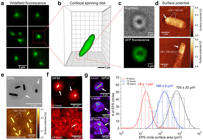
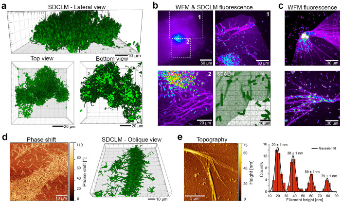
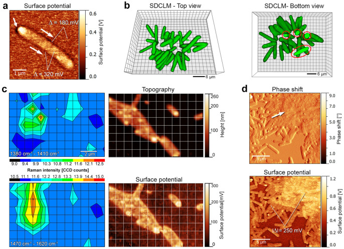
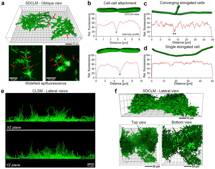
Microorganism pathogenicity strongly relies on the generation of multicellular assemblies, called biofilms. Understanding their organization can unveil vulnerabilities leading to potential treatments; spatially and temporally-resolved comprehensive experimental characterization can provide new details of biofilm formation, and possibly new targets for disease control. Here, biofilm formation of economically important phytopathogen Xylella fastidiosa was analyzed at single-cell resolution using nanometer-resolution spectro-microscopy techniques, addressing the role of different types of extracellular polymeric substances (EPS) at each stage of the entire bacterial life cycle. Single cell adhesion is caused by unspecific electrostatic interactions through proteins at the cell polar region, where EPS accumulation is required for more firmly-attached, irreversibly adhered cells. Subsequently, bacteria form clusters, which are embedded in secreted loosely-bound EPS, and bridged by up to ten-fold elongated cells that form the biofilm framework. During biofilm maturation, soluble EPS forms a filamentous matrix that facilitates cell adhesion and provides mechanical support, while the biofilm keeps anchored by few cells. This floating architecture maximizes nutrient distribution while allowing detachment upon larger shear stresses; it thus complies with biological requirements of the bacteria life cycle. Using new approaches, our findings provide insights regarding different aspects of the adhesion process of X. fastidiosa and biofilm formation.
微生物致病性在很大程度上依赖于称为生物膜的多细胞聚集体的形成。了解它们的组织结构可以揭示潜在治疗方法的薄弱环节;空间和时间分辨的全面实验表征可以提供生物膜形成的新细节,并可能为疾病控制提供新的靶点。在这里,使用纳米分辨率光谱显微镜技术在单细胞分辨率下分析了经济上重要的植物病原体——木质部难养菌的生物膜形成,探讨了不同类型的胞外聚合物(EPS)在整个细菌生命周期各阶段的作用。单细胞粘附是由细胞极性区域的蛋白质通过非特异性静电相互作用引起的,在该区域,EPS的积累对于更牢固附着、不可逆粘附的细胞是必需的。随后,细菌形成簇,这些簇嵌入分泌的松散结合的EPS中,并由形成生物膜框架的长达十倍伸长的细胞桥接。在生物膜成熟过程中,可溶性EPS形成丝状基质,促进细胞粘附并提供机械支撑,而生物膜由少数细胞保持锚定。这种漂浮结构在允许在较大剪切应力下脱离的同时最大限度地提高了营养物质的分布;因此它符合细菌生命周期的生物学要求。通过新方法,我们的研究结果提供了关于木质部难养菌粘附过程和生物膜形成不同方面的见解。