Wang Jing, Li Chun, Cao Yuan, Wang Qiyan, Lu Linghui, Chang Hong, Wu Yan, Han Jing, Wang Wei, Tu Pengfei, Wang Yong
Modern Research Center for Traditional Chinese Medicine, School of Chinese Materia Medica, Beijing University of Chinese Medicine, Beijing, China.
Basic Medical College, Beijing University of Chinese Medicine, Beijing, China.
BMC Complement Altern Med. 2015 Oct 7;15:352. doi: 10.1186/s12906-015-0869-z.
Qi-shen-yi-qi (QSYQ), one of the most well-known traditional Chinese medicine (TCM) formulas, has been shown to have cardioprotective effects in rats with heart failure (HF) induced by acute myocardial infarction (AMI). However, the mechanisms of its therapeutic effects remain unclear. In this study, we aim to explore the mechanisms of QSYQ in preventing left ventricular remodelling in rats with HF. The anti-apoptosis an anti-inflammation effects of QSYQ were investigated.
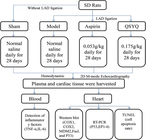
Sprague-Dawley (SD) rats were randomly divided into 4 groups: sham group, model group, QSYQ treatment group and aspirin group. Heart failure model was induced by ligation of left anterior descending (LAD) coronary artery. 28 days after surgery, hemodynamics were detected. Echocardiography was adopted to evaluate heart function. TUNEL assay was applied to assess myocardial apoptosis rates. Protein expressions of cyclooxygenase1 and 2 (COX1and COX2), Fas ligand (FasL), P53 and MDM2 were measured by western-blot. RT-PCR was applied to detect expressions of our subtype receptors of PGE2 (EP1, 2, 3, and 4).
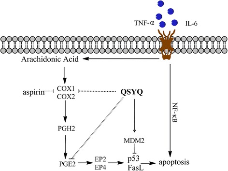
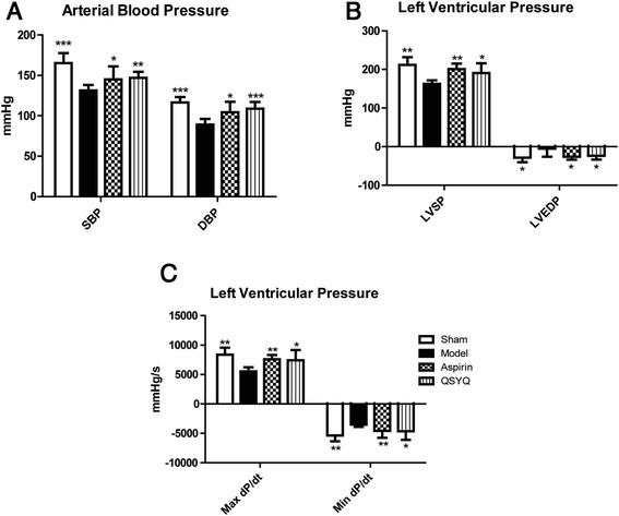
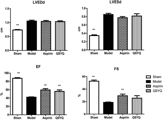
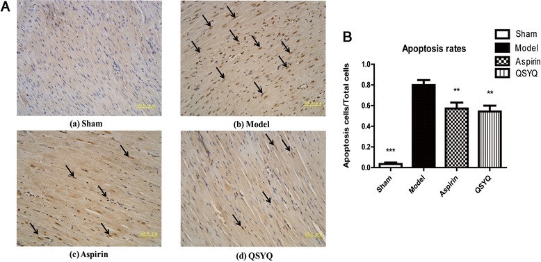
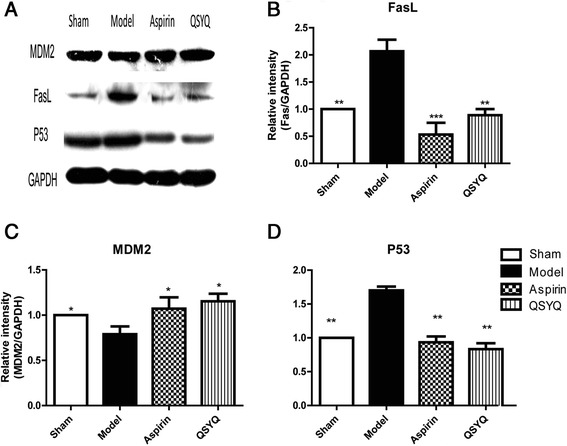
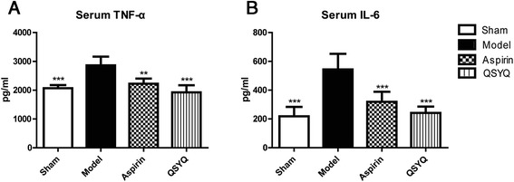
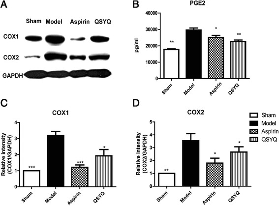
Ultrasonography showed that EF and FS values decreased significantly and abnormal hemodynamic alterations were observed in model group compared to sham group. These indications illustrated that HF models were successfully induced. Levels of inflammatory cytokines (TNF-α and IL-6) in myocardial tissue were up-regulated in the model group as compared to those in sham group. Western-blot analysis showed that cyclooxygenase 2, which is highly inducible by inflammatory cytokines, increased significantly. Moreover, RT-PCR showed that expressions of EP2 and EP4, which are the receptors of PGE2, were also up-regulated. Increased expressions of apoptotic pathway factors, including P53 and FasL, might be induced by the binding of PGE2 with EP2/4. MDM2, the inhibitor of P53, decreased in model group. TUNEL results manifested that apoptosis rates of myocardial cells increased in the model group. After treatment with QSYQ, expressions of inflammatory factors, including TNF-α, IL-6 and COX2, were reduced. Expressions of EP2 and EP4 receptors also decreased, suggesting that PGE2-mediated apoptosis was inhibited by QSYQ. MDM2 was up-regulated and P53 and FasL in the apoptotic pathway were down-regulated. Apoptosis rates in myocardial tissue in the QSYQ group decreased compared with those in the model group.
QSYQ exerts cardiac protective efficacy mainly through inhibiting the inflammatory response and down-regulating apoptosis. The anti-inflammatory and anti-apoptosis efficacies of QSYQ are probably achieved by inhibition of COXs-induced P53/FasL pathway. These findings provide experimental evidence for the beneficial effects of QSYQ in the clinical application for treating patients with HF.
芪参益气滴丸(QSYQ)是最著名的中药方剂之一,已被证明对急性心肌梗死(AMI)诱导的心力衰竭(HF)大鼠具有心脏保护作用。然而,其治疗作用的机制仍不清楚。在本研究中,我们旨在探讨QSYQ预防HF大鼠左心室重构的机制。研究了QSYQ的抗凋亡和抗炎作用。
将Sprague-Dawley(SD)大鼠随机分为4组:假手术组、模型组、QSYQ治疗组和阿司匹林组。通过结扎左冠状动脉前降支(LAD)诱导心力衰竭模型。术后28天,检测血流动力学。采用超声心动图评估心脏功能。应用TUNEL法评估心肌凋亡率。通过蛋白质印迹法检测环氧化酶1和2(COX1和COX2)、Fas配体(FasL)、P53和MDM2的蛋白表达。应用RT-PCR检测前列腺素E2(PGE2)各亚型受体(EP1、2、3和4)的表达。
超声检查显示,与假手术组相比,模型组的射血分数(EF)和缩短分数(FS)值显著降低,且观察到异常的血流动力学改变。这些指标表明成功诱导了HF模型。与假手术组相比,模型组心肌组织中炎性细胞因子(TNF-α和IL-6)水平上调。蛋白质印迹分析显示,可被炎性细胞因子高度诱导的环氧化酶2显著增加。此外,RT-PCR显示,作为PGE2受体的EP2和EP4的表达也上调。PGE2与EP2/4结合可能诱导凋亡途径因子(包括P53和FasL)表达增加。模型组中P53的抑制剂MDM2减少。TUNEL结果表明,模型组心肌细胞凋亡率增加。用QSYQ治疗后,包括TNF-α、IL-6和COX2在内的炎性因子表达降低。EP2和EP4受体的表达也降低,表明QSYQ抑制了PGE2介导的凋亡。MDM2上调,凋亡途径中的P53和FasL下调。与模型组相比,QSYQ组心肌组织中的凋亡率降低。
QSYQ主要通过抑制炎症反应和下调凋亡发挥心脏保护作用。QSYQ的抗炎和抗凋亡作用可能是通过抑制COXs诱导的P53/FasL途径实现的。这些发现为QSYQ在治疗HF患者的临床应用中的有益作用提供了实验证据。