School of Engineering and Applied Sciences, Wyss Institute for Biologically Inspired Engineering, Harvard University, Cambridge, Massachusetts, United States of America.
Pharmaceutical Sciences, Roche Pharma Research and Early Development, Roche Innovation Center, Basel, Switzerland.
Sci Rep. 2016 Oct 11;6:34845. doi: 10.1038/srep34845.
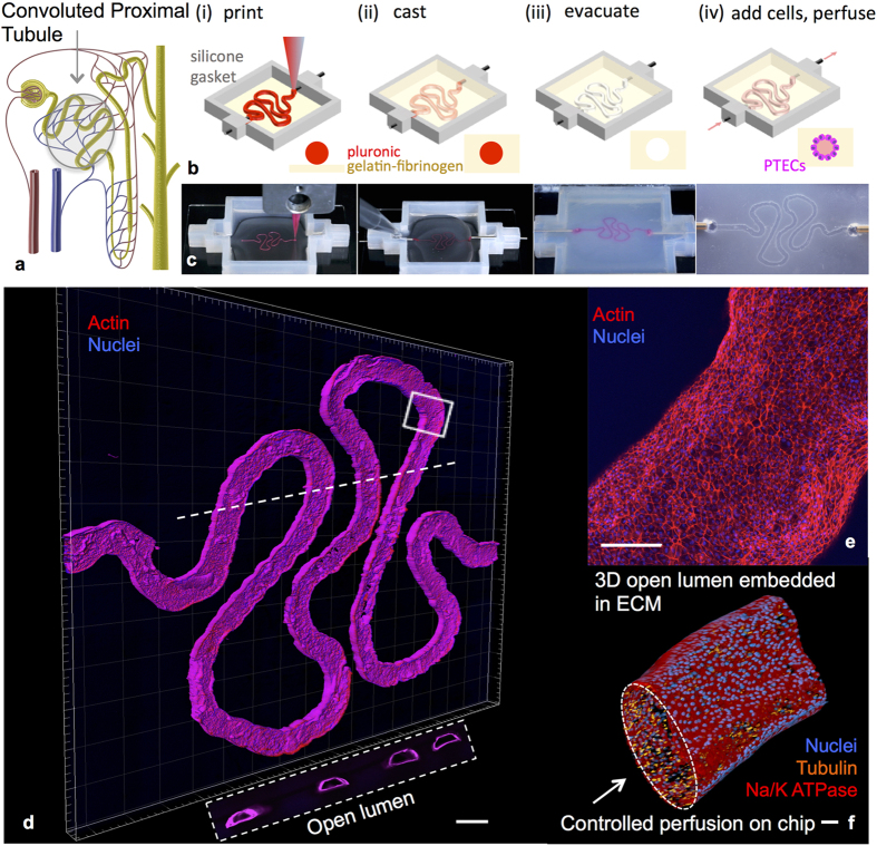
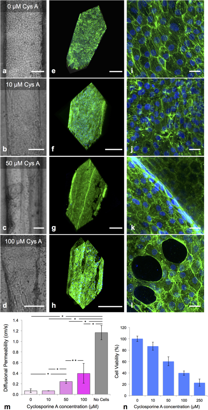
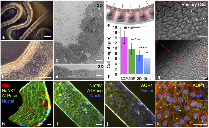
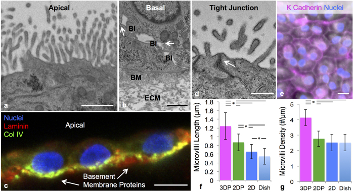
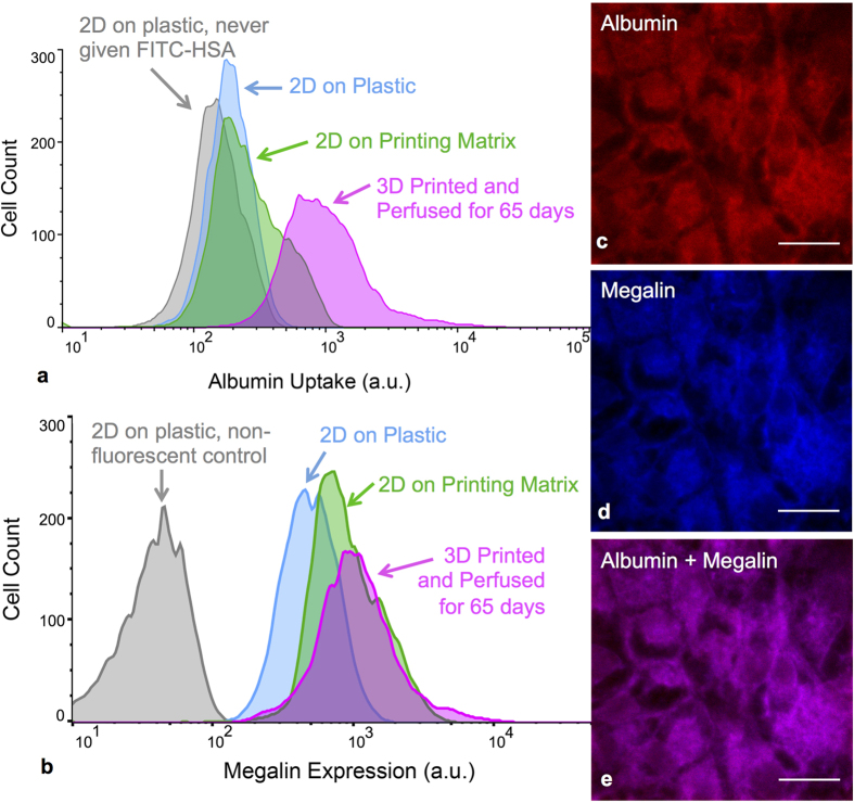
Three-dimensional models of kidney tissue that recapitulate human responses are needed for drug screening, disease modeling, and, ultimately, kidney organ engineering. Here, we report a bioprinting method for creating 3D human renal proximal tubules in vitro that are fully embedded within an extracellular matrix and housed in perfusable tissue chips, allowing them to be maintained for greater than two months. Their convoluted tubular architecture is circumscribed by proximal tubule epithelial cells and actively perfused through the open lumen. These engineered 3D proximal tubules on chip exhibit significantly enhanced epithelial morphology and functional properties relative to the same cells grown on 2D controls with or without perfusion. Upon introducing the nephrotoxin, Cyclosporine A, the epithelial barrier is disrupted in a dose-dependent manner. Our bioprinting method provides a new route for programmably fabricating advanced human kidney tissue models on demand.
需要能够重现人体反应的肾脏组织三维模型,以进行药物筛选、疾病建模,最终实现肾脏器官工程。在此,我们报告了一种生物打印方法,可用于体外创建完全嵌入细胞外基质并置于可灌注组织芯片中的三维人肾近端小管,从而可以将其维持超过两个月。这些曲折的管状结构由近端小管上皮细胞包围,并通过开放腔主动灌注。与在 2D 对照物上培养的具有或不具有灌注的相同细胞相比,这些在芯片上构建的工程化 3D 近端小管表现出明显增强的上皮形态和功能特性。当引入肾毒物环孢素 A 时,上皮屏障以剂量依赖性方式被破坏。我们的生物打印方法为按需程序化制造先进的人类肾脏组织模型提供了新途径。