• 文献检索
  • 文档翻译
  • 深度研究
  • 学术资讯
  • Suppr Zotero 插件Zotero 插件
  • 邀请有礼
  • 套餐&价格
  • 历史记录
应用&插件
Suppr Zotero 插件Zotero 插件浏览器插件Mac 客户端Windows 客户端微信小程序
定价
高级版会员购买积分包购买API积分包
服务
文献检索文档翻译深度研究API 文档MCP 服务
关于我们
关于 Suppr公司介绍联系我们用户协议隐私条款
关注我们

Suppr 超能文献

核心技术专利:CN118964589B侵权必究
粤ICP备2023148730 号-1Suppr @ 2026

文献检索

告别复杂PubMed语法,用中文像聊天一样搜索,搜遍4000万医学文献。AI智能推荐,让科研检索更轻松。

立即免费搜索

文件翻译

保留排版,准确专业,支持PDF/Word/PPT等文件格式,支持 12+语言互译。

免费翻译文档

深度研究

AI帮你快速写综述,25分钟生成高质量综述,智能提取关键信息,辅助科研写作。

立即免费体验

基于轮廓追踪与校正的胸部CT中含胸膜下结节的肺分割自动方法

Automatic Approach for Lung Segmentation with Juxta-Pleural Nodules from Thoracic CT Based on Contour Tracing and Correction.

作者信息

Wang Jinke, Guo Haoyan

机构信息

Department of Software Engineering, Harbin University of Science and Technology, Rongcheng, China.

School of Computer Science and Technology, Harbin Institute of Technology, Weihai, China.

出版信息

Comput Math Methods Med. 2016;2016:2962047. doi: 10.1155/2016/2962047. Epub 2016 Nov 16.

DOI:10.1155/2016/2962047
PMID:27974907
原文链接:https://pmc.ncbi.nlm.nih.gov/articles/PMC5128731/
Abstract

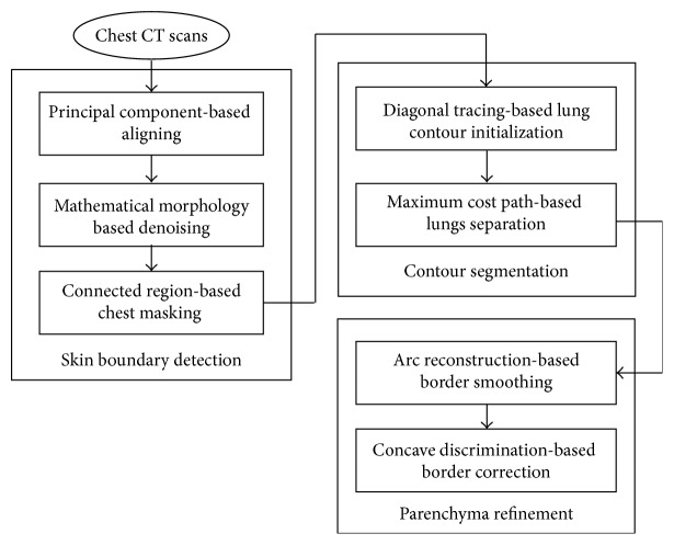
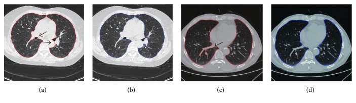
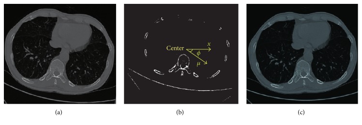
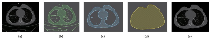
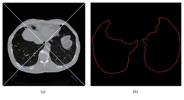
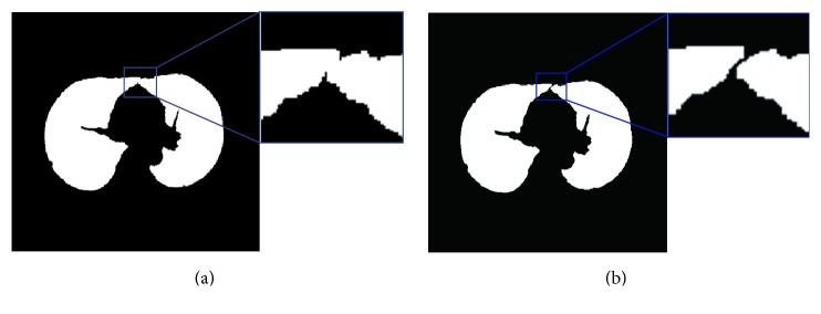
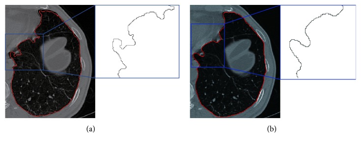
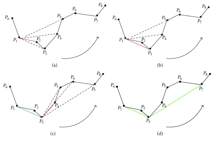
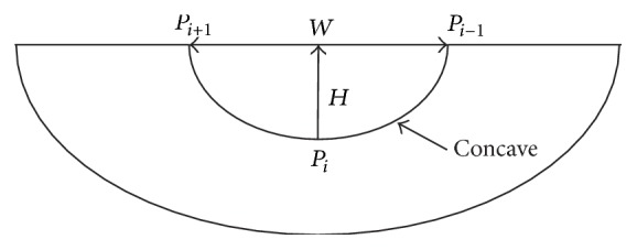
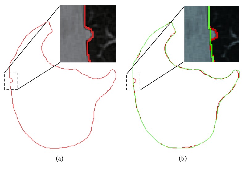
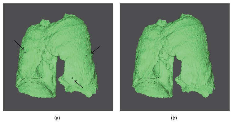
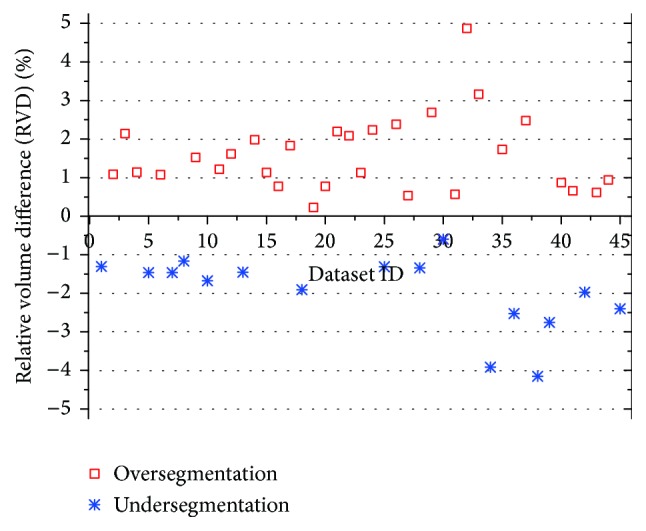
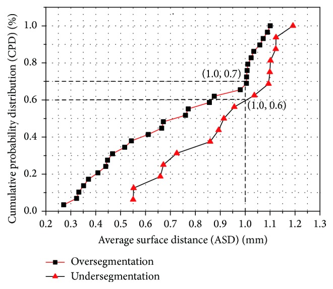
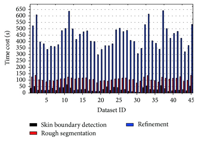
This paper presents a fully automatic framework for lung segmentation, in which juxta-pleural nodule problem is brought into strong focus. The proposed scheme consists of three phases: skin boundary detection, rough segmentation of lung contour, and pulmonary parenchyma refinement. Firstly, chest skin boundary is extracted through image aligning, morphology operation, and connective region analysis. Secondly, diagonal-based border tracing is implemented for lung contour segmentation, with maximum cost path algorithm used for separating the left and right lungs. Finally, by arc-based border smoothing and concave-based border correction, the refined pulmonary parenchyma is obtained. The proposed scheme is evaluated on 45 volumes of chest scans, with volume difference (VD) 11.15 ± 69.63 cm, volume overlap error (VOE) 3.5057 ± 1.3719%, average surface distance (ASD) 0.7917 ± 0.2741 mm, root mean square distance (RMSD) 1.6957 ± 0.6568 mm, maximum symmetric absolute surface distance (MSD) 21.3430 ± 8.1743 mm, and average time-cost 2 seconds per image. The preliminary results on accuracy and complexity prove that our scheme is a promising tool for lung segmentation with juxta-pleural nodules.

摘要

本文提出了一种用于肺部分割的全自动框架,其中胸膜旁结节问题得到了重点关注。所提出的方案包括三个阶段:皮肤边界检测、肺轮廓的粗略分割以及肺实质细化。首先,通过图像对齐、形态学操作和连通区域分析来提取胸部皮肤边界。其次,采用基于对角线的边界追踪进行肺轮廓分割,并使用最大代价路径算法来分离左右肺。最后,通过基于弧线的边界平滑和基于凹面的边界校正,获得细化的肺实质。该方案在45例胸部扫描图像上进行了评估,体积差异(VD)为11.15±69.63cm,体积重叠误差(VOE)为3.5057±1.3719%,平均表面距离(ASD)为0.7917±0.2741mm,均方根距离(RMSD)为1.6957±0.6568mm,最大对称绝对表面距离(MSD)为21.3430±8.1743mm,平均每张图像的时间成本为2秒。在准确性和复杂性方面的初步结果证明,我们的方案是一种用于分割伴有胸膜旁结节的肺部的有前景的工具。

https://cdn.ncbi.nlm.nih.gov/pmc/blobs/6d4e/5128731/4cba2c0da031/CMMM2016-2962047.016.jpg
https://cdn.ncbi.nlm.nih.gov/pmc/blobs/6d4e/5128731/21a7fad4ca8e/CMMM2016-2962047.001.jpg
https://cdn.ncbi.nlm.nih.gov/pmc/blobs/6d4e/5128731/527d10b94739/CMMM2016-2962047.002.jpg
https://cdn.ncbi.nlm.nih.gov/pmc/blobs/6d4e/5128731/7ccf9bac919d/CMMM2016-2962047.003.jpg
https://cdn.ncbi.nlm.nih.gov/pmc/blobs/6d4e/5128731/171cb4c7ad52/CMMM2016-2962047.004.jpg
https://cdn.ncbi.nlm.nih.gov/pmc/blobs/6d4e/5128731/c77ecac9ea76/CMMM2016-2962047.005.jpg
https://cdn.ncbi.nlm.nih.gov/pmc/blobs/6d4e/5128731/e91685b19049/CMMM2016-2962047.006.jpg
https://cdn.ncbi.nlm.nih.gov/pmc/blobs/6d4e/5128731/28bcb800202a/CMMM2016-2962047.007.jpg
https://cdn.ncbi.nlm.nih.gov/pmc/blobs/6d4e/5128731/be1085b3d218/CMMM2016-2962047.008.jpg
https://cdn.ncbi.nlm.nih.gov/pmc/blobs/6d4e/5128731/0cfe156cdfe7/CMMM2016-2962047.009.jpg
https://cdn.ncbi.nlm.nih.gov/pmc/blobs/6d4e/5128731/884534b3bd7d/CMMM2016-2962047.010.jpg
https://cdn.ncbi.nlm.nih.gov/pmc/blobs/6d4e/5128731/3a40043f9532/CMMM2016-2962047.011.jpg
https://cdn.ncbi.nlm.nih.gov/pmc/blobs/6d4e/5128731/18e4aa030808/CMMM2016-2962047.012.jpg
https://cdn.ncbi.nlm.nih.gov/pmc/blobs/6d4e/5128731/46bced0fd331/CMMM2016-2962047.013.jpg
https://cdn.ncbi.nlm.nih.gov/pmc/blobs/6d4e/5128731/437396f90be4/CMMM2016-2962047.014.jpg
https://cdn.ncbi.nlm.nih.gov/pmc/blobs/6d4e/5128731/a960883ae5fa/CMMM2016-2962047.015.jpg
https://cdn.ncbi.nlm.nih.gov/pmc/blobs/6d4e/5128731/4cba2c0da031/CMMM2016-2962047.016.jpg

相似文献

1
Automatic Approach for Lung Segmentation with Juxta-Pleural Nodules from Thoracic CT Based on Contour Tracing and Correction.基于轮廓追踪与校正的胸部CT中含胸膜下结节的肺分割自动方法
Comput Math Methods Med. 2016;2016:2962047. doi: 10.1155/2016/2962047. Epub 2016 Nov 16.
2
Segmentation of pulmonary nodules using adaptive local region energy with probability density function-based similarity distance and multi-features clustering.基于概率密度函数相似性距离和多特征聚类的自适应局部区域能量法用于肺结节分割
Biomed Eng Online. 2016 May 5;15(1):49. doi: 10.1186/s12938-016-0164-3.
3
Fully automatic deep learning-based lung parenchyma segmentation and boundary correction in thoracic CT scans.基于深度学习的全自动化肺部组织分割和胸 CT 扫描边界修正。
Int J Comput Assist Radiol Surg. 2024 Feb;19(2):261-272. doi: 10.1007/s11548-023-03010-0. Epub 2023 Aug 18.
4
Smoothing lung segmentation surfaces in three-dimensional X-ray CT images using anatomic guidance.利用解剖学引导在三维X射线CT图像中平滑肺部分割表面
Acad Radiol. 2005 Dec;12(12):1502-11. doi: 10.1016/j.acra.2005.08.008.
5
A fully automatic segmentation algorithm for CT lung images based on random forest.基于随机森林的 CT 肺图像全自动分割算法。
Med Phys. 2020 Feb;47(2):518-529. doi: 10.1002/mp.13939. Epub 2019 Dec 29.
6
Juxta-Vascular Pulmonary Nodule Segmentation in PET-CT Imaging Based on an LBF Active Contour Model with Information Entropy and Joint Vector.基于具有信息熵和联合向量的LBF主动轮廓模型的PET-CT成像中的血管旁肺结节分割
Comput Math Methods Med. 2018 Jan 8;2018:2183847. doi: 10.1155/2018/2183847. eCollection 2018.
7
Automatic Lung Segmentation With Juxta-Pleural Nodule Identification Using Active Contour Model and Bayesian Approach.基于主动轮廓模型和贝叶斯方法的具有胸膜旁结节识别功能的自动肺分割
IEEE J Transl Eng Health Med. 2018 May 18;6:1800513. doi: 10.1109/JTEHM.2018.2837901. eCollection 2018.
8
Automated lung segmentation in digital chest tomosynthesis.数字断层合成胸部 CT 中的自动肺分割。
Med Phys. 2012 Feb;39(2):732-41. doi: 10.1118/1.3671939.
9
Fuzzy speed function based active contour model for segmentation of pulmonary nodules.基于模糊速度函数的主动轮廓模型用于肺结节分割
Biomed Mater Eng. 2014;24(1):539-47. doi: 10.3233/BME-130840.
10
Automated iterative neutrosophic lung segmentation for image analysis in thoracic computed tomography.自动化迭代 Neutrosophic 肺分割用于胸部 CT 图像分析。
Med Phys. 2013 Aug;40(8):081912. doi: 10.1118/1.4812679.

引用本文的文献

1
Tumor segmentation via enhanced area growth algorithm for lung CT images.基于改进区域生长算法的肺部 CT 图像肿瘤分割。
BMC Med Imaging. 2023 Nov 20;23(1):189. doi: 10.1186/s12880-023-01126-y.
2
[Corona virus disease 2019 lesion segmentation network based on an adaptive joint loss function].基于自适应联合损失函数的2019冠状病毒病病变分割网络
Sheng Wu Yi Xue Gong Cheng Xue Za Zhi. 2023 Aug 25;40(4):743-752. doi: 10.7507/1001-5515.202206051.
3
Generalization optimizing machine learning to improve CT scan radiomics and assess immune checkpoint inhibitors' response in non-small cell lung cancer: a multicenter cohort study.

本文引用的文献

1
Contour propagation using feature-based deformable registration for lung cancer.使用基于特征的可变形配准进行肺癌轮廓传播
Biomed Res Int. 2013;2013:701514. doi: 10.1155/2013/701514. Epub 2013 Dec 2.
2
Multi-stage learning for robust lung segmentation in challenging CT volumes.用于在具有挑战性的CT容积数据中进行稳健肺部分割的多阶段学习。
Med Image Comput Comput Assist Interv. 2011;14(Pt 3):667-74. doi: 10.1007/978-3-642-23626-6_82.
3
Automated 3-D segmentation of lungs with lung cancer in CT data using a novel robust active shape model approach.
优化机器学习以改善CT扫描影像组学并评估免疫检查点抑制剂在非小细胞肺癌中的反应:一项多中心队列研究
Front Oncol. 2023 Jul 20;13:1196414. doi: 10.3389/fonc.2023.1196414. eCollection 2023.
4
A Systematic Review of Automated Segmentation Methods and Public Datasets for the Lung and its Lobes and Findings on Computed Tomography Images.基于 CT 图像的肺部及其叶部自动分割方法和公共数据集的系统评价及相关发现。
Yearb Med Inform. 2022 Aug;31(1):277-295. doi: 10.1055/s-0042-1742517. Epub 2022 Dec 4.
5
Automatic Lung Segmentation With Juxta-Pleural Nodule Identification Using Active Contour Model and Bayesian Approach.基于主动轮廓模型和贝叶斯方法的具有胸膜旁结节识别功能的自动肺分割
IEEE J Transl Eng Health Med. 2018 May 18;6:1800513. doi: 10.1109/JTEHM.2018.2837901. eCollection 2018.
利用一种新颖的稳健主动形状模型方法对 CT 数据中的肺癌进行自动三维肺分割。
IEEE Trans Med Imaging. 2012 Feb;31(2):449-60. doi: 10.1109/TMI.2011.2171357. Epub 2011 Oct 13.
4
Automatic lung segmentation from thoracic computed tomography scans using a hybrid approach with error detection.使用带有错误检测的混合方法从胸部计算机断层扫描中自动进行肺部分割。
Med Phys. 2009 Jul;36(7):2934-47. doi: 10.1118/1.3147146.
5
Texture classification-based segmentation of lung affected by interstitial pneumonia in high-resolution CT.基于纹理分类的高分辨率CT中受间质性肺炎影响的肺部分割
Med Phys. 2008 Dec;35(12):5290-302. doi: 10.1118/1.3003066.
6
Automatic segmentation of lung parenchyma in the presence of diseases based on curvature of ribs.基于肋骨曲率的疾病状态下肺实质自动分割
Acad Radiol. 2008 Sep;15(9):1173-80. doi: 10.1016/j.acra.2008.02.004.
7
Correction of segmented lung boundary for inclusion of pleural nodules and pulmonary vessels in chest CT images.胸部CT图像中用于纳入胸膜结节和肺血管的分割肺边界校正
Comput Biol Med. 2008 Aug;38(8):845-57. doi: 10.1016/j.compbiomed.2008.04.012. Epub 2008 Jun 26.
8
Adaptive border marching algorithm: automatic lung segmentation on chest CT images.自适应边界行进算法:胸部CT图像上的自动肺部分割
Comput Med Imaging Graph. 2008 Sep;32(6):452-62. doi: 10.1016/j.compmedimag.2008.04.005. Epub 2008 Jun 2.
9
3D computerized segmentation of lung volume with computed tomography.利用计算机断层扫描进行肺容积的三维计算机分割
Acad Radiol. 2006 Jun;13(6):670-7. doi: 10.1016/j.acra.2006.02.039.
10
Smoothing lung segmentation surfaces in three-dimensional X-ray CT images using anatomic guidance.利用解剖学引导在三维X射线CT图像中平滑肺部分割表面
Acad Radiol. 2005 Dec;12(12):1502-11. doi: 10.1016/j.acra.2005.08.008.