Graduate Institute of Opto-Mechatronics, National Chung Cheng University, Chia-Yi 621, Taiwan.
Department of Mechanical Engineering, National Chung Cheng University, Chia-Yi 621, Taiwan.
Sensors (Basel). 2017 May 6;17(5):1053. doi: 10.3390/s17051053.
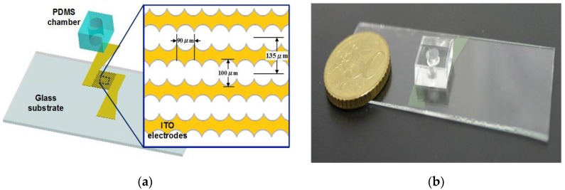
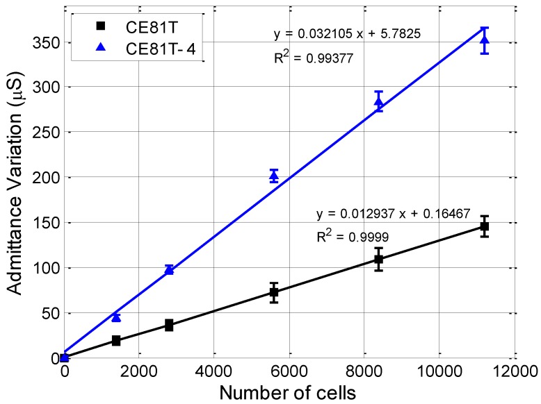
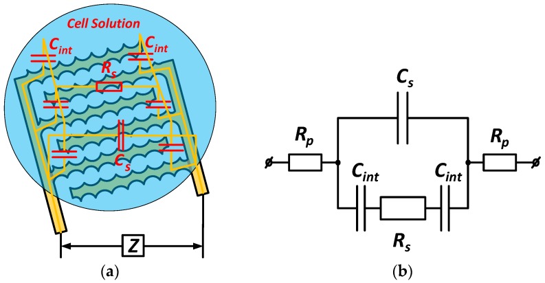
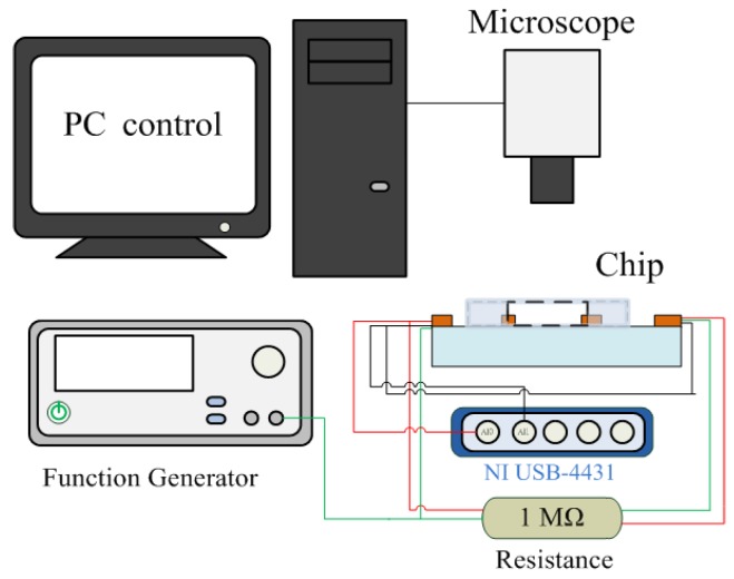
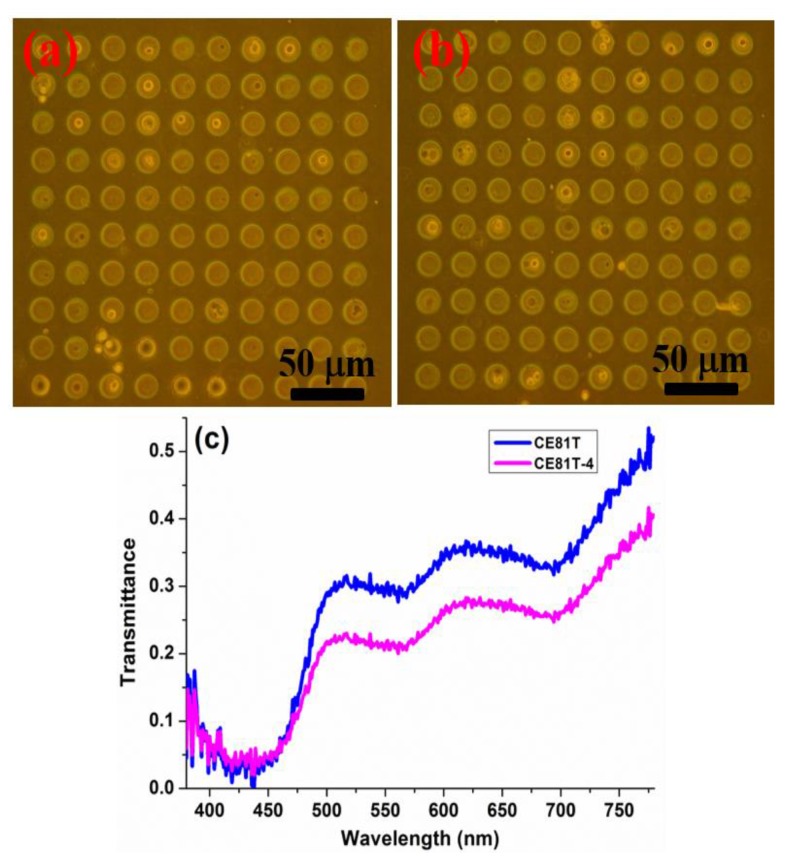
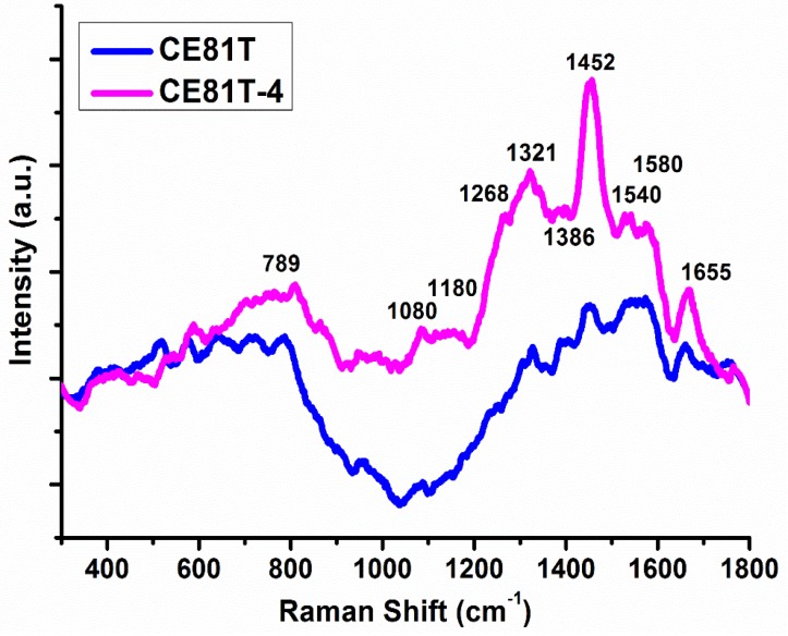
Analysis of cancerous cells allows us to provide useful information for the early diagnosis of cancer and to monitor treatment progress. An approach based on electrical principles has recently become an attractive technique. This study presents a microdevice that utilizes a dielectrophoretic impedance measurement method for the identification of cancerous cells. The proposed biochip consists of circle-on-line microelectrodes that are patterned using a standard microfabrication processes. A sample of various cell concentrations was introduced in an open-top microchamber. The target cells were collectively concentrated between the microelectrodes using dielectrophoresis manipulation, and their electrical impedance properties were also measured. Different stages of human esophageal squamous cell carcinoma lines could be distinguished. This result is consistent with findings using hyperspectral imaging technology. Moreover, it was observed that the distinguishing characteristics change in response to the progression of cancer cell invasiveness by Raman spectroscopy. The device enables highly efficient cell collection and provides rapid, sensitive, and label-free electrical measurements of cancerous cells.
癌细胞分析可提供有助于癌症早期诊断和监测治疗进展的有用信息。基于电学原理的方法最近成为一种有吸引力的技术。本研究提出了一种利用介电泳阻抗测量方法识别癌细胞的微器件。所提出的生物芯片由圆形在线微电极组成,这些微电极采用标准微加工工艺进行图案化。将不同细胞浓度的样本引入开顶微腔中。使用介电泳操作将靶细胞集中在微电极之间,并测量其电阻抗特性。可以区分不同阶段的人食管鳞状细胞癌细胞系。这一结果与使用高光谱成像技术的结果一致。此外,还通过拉曼光谱观察到,区分特征随癌细胞侵袭性的进展而变化。该装置能够高效地收集细胞,并对癌细胞进行快速、灵敏且无需标记的电阻抗测量。