Barkai Omer, Goldstein Robert H, Caspi Yaki, Katz Ben, Lev Shaya, Binshtok Alexander M
Department of Medical Neurobiology, Institute for Medical Research Israel-Canada, Hadassah School of Medicine, The Hebrew University-Hadassah School of MedicineJerusalem, Israel.
The Edmond and Lily Safra Center for Brain Sciences, The Hebrew University of JerusalemJerusalem, Israel.
Front Mol Neurosci. 2017 Jun 13;10:181. doi: 10.3389/fnmol.2017.00181. eCollection 2017.
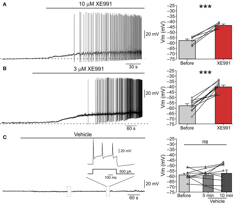
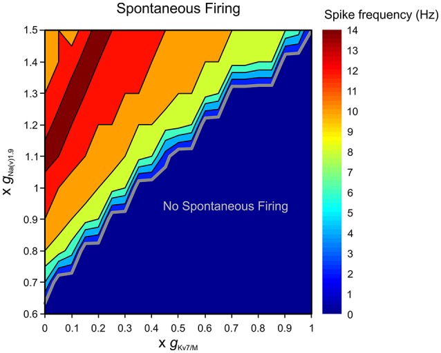
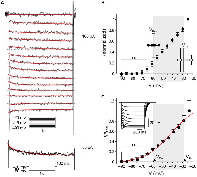
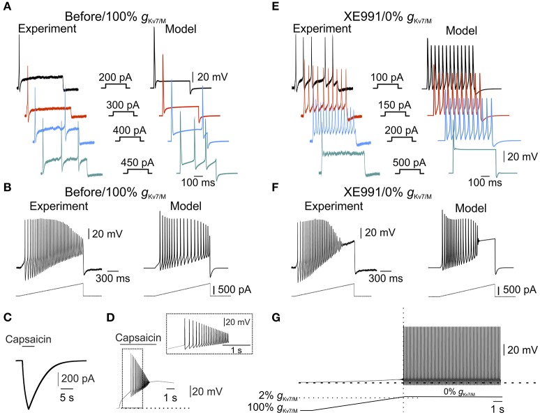
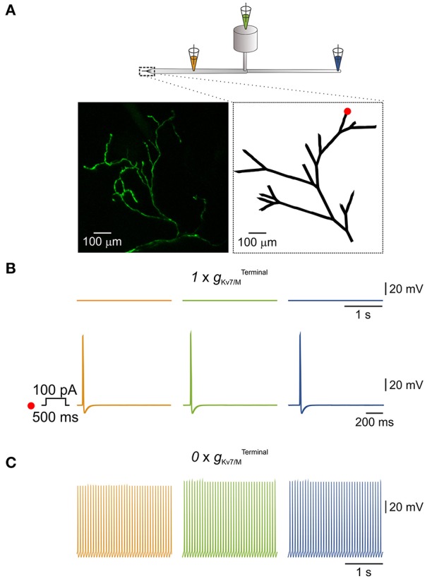
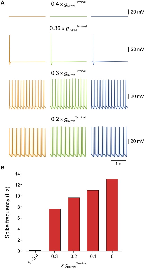
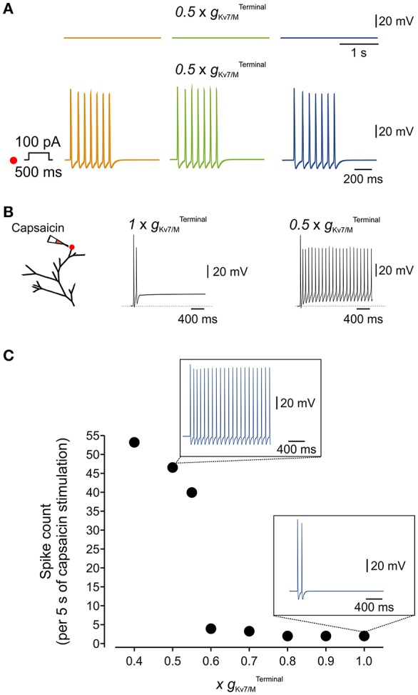
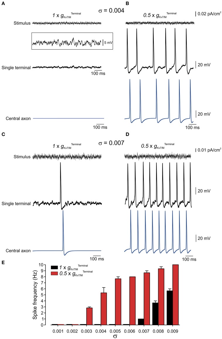
Peripheral nociceptive neurons encode and convey injury-inducing stimuli toward the central nervous system. In normal conditions, tight control of nociceptive resting potential prevents their spontaneous activation. However, in many pathological conditions the control of membrane potential is disrupted, leading to ectopic, stimulus-unrelated firing of nociceptive neurons, which is correlated to spontaneous pain. We have investigated the role of K7/M channels in stabilizing membrane potential and impeding spontaneous firing of nociceptive neurons. These channels generate low voltage-activating, noninactivating M-type K currents (M-current, ), which control neuronal excitability. Using perforated-patch recordings from cultured, rat nociceptor-like dorsal root ganglion neurons, we show that inhibition of M-current leads to depolarization of nociceptive neurons and generation of repetitive firing. To assess to what extent the M-current, acting at the nociceptive terminals, is able to stabilize terminals' membrane potential, thus preventing their ectopic activation, in normal and pathological conditions, we built a multi-compartment computational model of a pseudo-unipolar unmyelinated nociceptive neuron with a realistic terminal tree. The modeled terminal tree was based on the structure of nociceptive peripheral terminal, which we assessed by multiphoton imaging of GFP-expressing nociceptive neuronal terminals innervating mice hind paw. By modifying the conductance of the K7/M channels at the modeled terminal tree (terminal g) we have found that 40% of the terminal g conductance is sufficient to prevent spontaneous firing, while ~75% of terminal g is sufficient to inhibit stimulus induced activation of nociceptive neurons. Moreover, we showed that terminal M-current reduces susceptibility of nociceptive neurons to a small fluctuations of membrane potentials. Furthermore, we simulated how the interaction between terminal persistent sodium current and M-current affects the excitability of the neurons. We demonstrated that terminal M-current in nociceptive neurons impeded spontaneous firing even when terminal Na1.9 channels conductance was substantially increased. On the other hand, when terminal g was decreased, nociceptive neurons fire spontaneously after slight increase in terminal Na1.9 conductance. Our results emphasize the pivotal role of M-current in stabilizing membrane potential and hereby in controlling nociceptive spontaneous firing, in normal and pathological conditions.
外周伤害性神经元编码并向中枢神经系统传递损伤诱导刺激。在正常情况下,对伤害性静息电位的严格控制可防止其自发激活。然而,在许多病理状态下,膜电位的控制被破坏,导致伤害性神经元出现异位的、与刺激无关的放电,这与自发痛相关。我们研究了K7/M通道在稳定膜电位和阻止伤害性神经元自发放电中的作用。这些通道产生低电压激活、非失活的M型钾电流(M电流),其控制神经元兴奋性。使用来自培养的大鼠伤害感受器样背根神经节神经元的穿孔膜片钳记录,我们发现抑制M电流会导致伤害性神经元去极化并产生重复放电。为了评估在正常和病理条件下,作用于伤害性终末的M电流在多大程度上能够稳定终末膜电位,从而防止其异位激活,我们构建了一个具有逼真终末树的假单极无髓鞘伤害性神经元的多室计算模型。建模的终末树基于伤害性外周终末的结构,我们通过对支配小鼠后爪的表达绿色荧光蛋白的伤害性神经元终末进行多光子成像来评估该结构。通过改变建模终末树(终末电导)处K7/M通道的电导,我们发现40%的终末电导足以防止自发放电,而约75%的终末电导足以抑制刺激诱导的伤害性神经元激活。此外,我们表明终末M电流降低了伤害性神经元对膜电位小波动的敏感性。此外,我们模拟了终末持续性钠电流与M电流之间的相互作用如何影响神经元的兴奋性。我们证明,即使终末Na1.9通道电导大幅增加,伤害性神经元中的终末M电流仍能阻止自发放电。另一方面,当终末电导降低时,终末Na1.9电导稍有增加,伤害性神经元就会自发放电。我们的结果强调了M电流在正常和病理条件下稳定膜电位以及由此控制伤害性自发放电中的关键作用。