School of Medicine, University of St Andrews, St Andrews, KY16 9TF, UK.
SUPA, School of Physics and Astronomy, University of St Andrews, KY16 9SS, St Andrews, UK.
Sci Rep. 2017 Aug 29;7(1):9844. doi: 10.1038/s41598-017-10234-z.
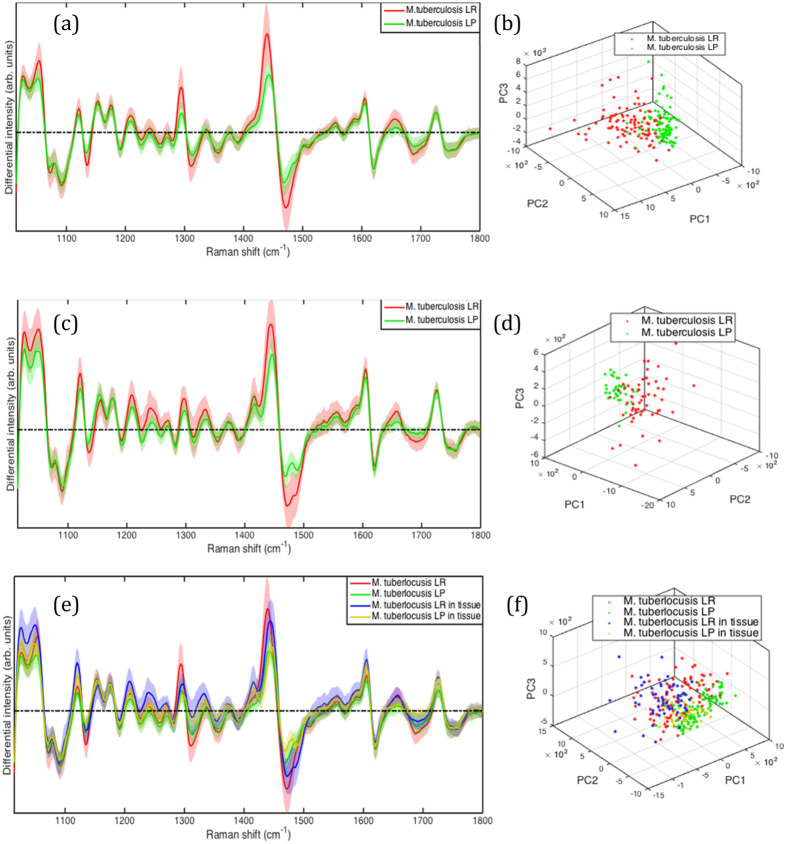
Tuberculosis relapse is a barrier to shorter treatment. It is thought that lipid rich cells, phenotypically resistant to antibiotics, may play a major role. Most studies investigating relapse use sputum samples although tissue bacteria may play an important role. We developed a non-destructive, label-free technique combining wavelength modulated Raman (WMR) spectroscopy and fluorescence detection (Nile Red staining) to interrogate Mycobacterium tuberculosis cell state. This approach could differentiate single "dormant" (lipid rich, LR) and "non-dormant" (lipid poor, LP) cells with high sensitivity and specificity. We applied this to experimentally infected guinea pig lung sections and were able to distinguish both cell types showing that the LR phenotype dominates in infected tissue. Both in-vitro and ex-vivo spectra correlated well, showing for the first time that Mycobacterium tuberculosis, likely to be phenotypically resistant to antibiotics, are present in large numbers in tissue. This is an important step in understanding the pathology of relapse supporting the idea that they may be caused by M. tuberculosis cells with lipid inclusions.
结核病复发是缩短治疗时间的障碍。人们认为富含脂类、表型上对抗生素耐药的细胞可能起主要作用。尽管组织细菌可能发挥重要作用,但大多数研究复发的研究都使用痰样本。我们开发了一种非破坏性、无标记的技术,结合波长调制拉曼(WMR)光谱和荧光检测(Nile Red 染色)来检测分枝杆菌细胞状态。这种方法可以高度敏感和特异性地区分单个“休眠”(富含脂类,LR)和“非休眠”(贫脂类,LP)细胞。我们将其应用于实验性感染豚鼠肺切片,能够区分这两种细胞类型,表明 LR 表型在感染组织中占主导地位。体内和体外的光谱都很好地相关,这是首次表明可能对抗生素表型耐药的分枝杆菌大量存在于组织中。这是了解复发病理学的重要一步,支持这样一种观点,即它们可能是由含有脂滴的分枝杆菌细胞引起的。