UCLA Cardiac Arrhythmia Center, UCLA Health System, David Geffen School of Medicine at UCLA, Los Angeles, California.
UCLA Department of Radiology, UCLA Health System, David Geffen School of Medicine at UCLA, Los Angeles, California.
Heart Rhythm. 2018 Feb;15(2):218-225. doi: 10.1016/j.hrthm.2017.10.003. Epub 2017 Oct 7.
Magnetic resonance imaging (MRI) has been performed safely in patients without MRI-conditional cardiac implantable electronic devices (CIEDs), but experience specifically with cardiac magnetic resonance imaging (CMR) is limited in this patient population.
Evaluate the safety of CMR in non-MRI-conditional CIEDs and the interpretability of images using wideband sequences.
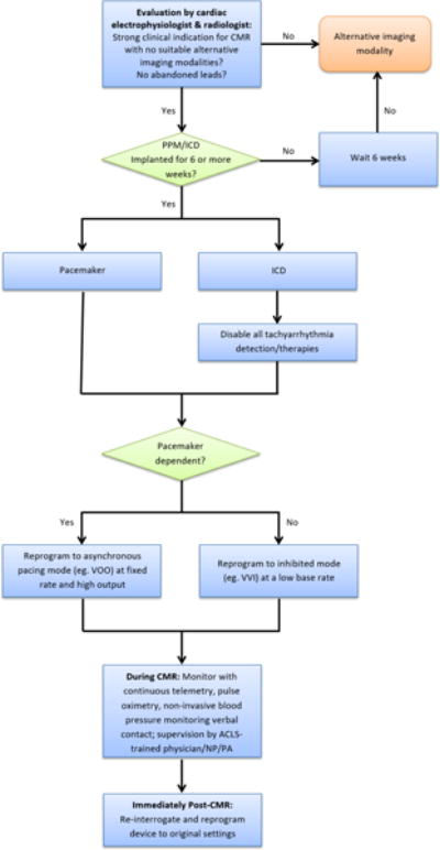
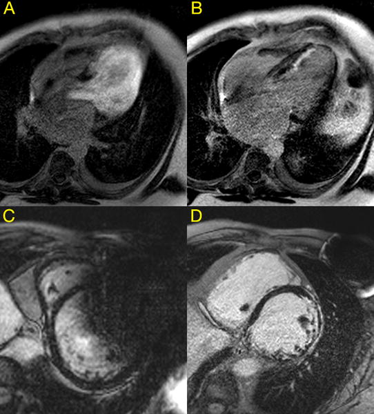
We performed 114 consecutive CMR studies in 111 patients (mean age 59 ± 14 years, with 12 pacemakers, 73 implantable cardioverter defibrillators, 29 biventricular defibrillators) using a wideband pulse sequence for late gadolinium enhancement (LGE) imaging. A standardized protocol for device management and patient monitoring was followed. Patients were evaluated for major clinical adverse events and device parameter changes immediately after CMR and at clinical follow-up.
In total, 111 CMR studies were completed successfully. There were no patient deaths, new arrhythmias, immediate generator or lead failures, electrical resets, or pacing capture failures in dependent patients. Right atrial, right ventricular, and left ventricular lead impedances were significantly lower post CMR, with median differences -7 Ω (interquartile range [IQR] -20 to 0 Ω; P < .0001), 0 Ω (IQR -19 to 0 Ω; P = .0001), and -10 Ω (IQR -30 to 0 Ω; P = .023), respectively. These changes persisted through the follow-up period, with median differences -18.5 Ω (IQR -41 to -66 Ω; P = .007), -19 Ω (IQR -44 to -7 Ω; P = .006), and -30 Ω (IQR -130 to 0 Ω; P = .003), respectively. Ninety-seven studies (87%) had no artifact limiting interpretation.
CMR can be performed safely in non-MRI-conditional CIEDs using a standardized protocol. Use of a wideband pulse sequence for LGE imaging yields a high rate of studies unaffected by artifact.
磁共振成像(MRI)已在无 MRI 条件的心脏植入式电子设备(CIED)患者中安全实施,但在该患者人群中,心脏磁共振成像(CMR)的经验有限。
评估非 MRI 条件的 CIED 进行 CMR 的安全性和宽带序列成像的可解读性。
我们使用宽带脉冲序列对 111 例(平均年龄 59±14 岁,包括 12 个起搏器、73 个植入式心脏复律除颤器、29 个双心室除颤器)连续进行了 114 例 CMR 研究,用于迟发钆增强(LGE)成像。遵循设备管理和患者监测的标准化方案。在 CMR 后和临床随访时评估患者主要临床不良事件和设备参数变化。
共有 111 例 CMR 研究成功完成。没有患者死亡、新出现的心律失常、立即发生的发生器或导联故障、电重置或依赖患者的起搏捕获失败。右心房、右心室和左心室导联阻抗在 CMR 后显著降低,中位数差异分别为-7Ω(四分位距 [IQR] -20 至 0Ω;P<.0001)、0Ω(IQR -19 至 0Ω;P=.0001)和-10Ω(IQR -30 至 0Ω;P=.023)。这些变化在随访期间持续存在,中位数差异分别为-18.5Ω(IQR -41 至 -66Ω;P=.007)、-19Ω(IQR -44 至 -7Ω;P=.006)和-30Ω(IQR -130 至 0Ω;P=.003)。97 项研究(87%)无影响解读的伪影。
使用标准化方案,非 MRI 条件的 CIED 可安全进行 CMR。宽带脉冲序列用于 LGE 成像可产生高比例不受伪影影响的研究。