Wang Xinggang, Yang Wei, Weinreb Jeffrey, Han Juan, Li Qiubai, Kong Xiangchuang, Yan Yongluan, Ke Zan, Luo Bo, Liu Tao, Wang Liang
Department of Radiology, Tongji Hospital, Huazhong University of Science and Technology, Jiefang Road 1095, 430030, Wuhan, China.
School of Electronics Information and Communications, Huazhong University of Science and Technology, Luoyu Road 1037, Wuhan, Hubei, 430074, China.
Sci Rep. 2017 Nov 13;7(1):15415. doi: 10.1038/s41598-017-15720-y.
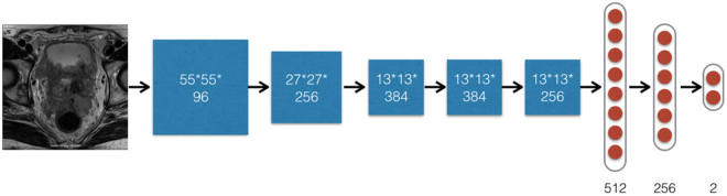
Prostate cancer (PCa) is a major cause of death since ancient time documented in Egyptian Ptolemaic mummy imaging. PCa detection is critical to personalized medicine and varies considerably under an MRI scan. 172 patients with 2,602 morphologic images (axial 2D T2-weighted imaging) of the prostate were obtained. A deep learning with deep convolutional neural network (DCNN) and a non-deep learning with SIFT image feature and bag-of-word (BoW), a representative method for image recognition and analysis, were used to distinguish pathologically confirmed PCa patients from prostate benign conditions (BCs) patients with prostatitis or prostate benign hyperplasia (BPH). In fully automated detection of PCa patients, deep learning had a statistically higher area under the receiver operating characteristics curve (AUC) than non-deep learning (P = 0.0007 < 0.001). The AUCs were 0.84 (95% CI 0.78-0.89) for deep learning method and 0.70 (95% CI 0.63-0.77) for non-deep learning method, respectively. Our results suggest that deep learning with DCNN is superior to non-deep learning with SIFT image feature and BoW model for fully automated PCa patients differentiation from prostate BCs patients. Our deep learning method is extensible to image modalities such as MR imaging, CT and PET of other organs.
前列腺癌(PCa)自古以来就是主要死因,埃及托勒密王朝木乃伊成像中有相关记载。PCa检测对个性化医疗至关重要,在MRI扫描下差异很大。我们获取了172例患者的2602张前列腺形态学图像(轴向二维T2加权成像)。使用深度卷积神经网络(DCNN)的深度学习方法以及具有尺度不变特征变换(SIFT)图像特征和词袋模型(BoW)的非深度学习方法(一种用于图像识别和分析的代表性方法),来区分经病理证实的PCa患者与患有前列腺炎或前列腺良性增生(BPH)的前列腺良性疾病(BCs)患者。在全自动检测PCa患者时,深度学习在受试者工作特征曲线(AUC)下的面积在统计学上高于非深度学习(P = 0.0007 < 0.001)。深度学习方法的AUC为0.84(95%置信区间0.78 - 0.89),非深度学习方法的AUC为0.70(95%置信区间0.63 - 0.77)。我们的结果表明,对于将PCa患者与前列腺BCs患者进行全自动区分,使用DCNN的深度学习优于使用SIFT图像特征和BoW模型的非深度学习。我们的深度学习方法可扩展到其他器官的MR成像、CT和PET等图像模态。