From the Department of Physiology, Anatomy, and Genetics (A.J.M.L., J.J.M., M.K.C., C.A.C., D.J.T.), Department of Physics, Clarendon Laboratory (J.J.M.), Radcliffe Department of Medicine (A.J.M.L., O.J.R., R.P.C., S.N.), and Acute Vascular Imaging Centre (R.P.C.), Radcliffe Department of Medicine, University of Oxford, United Kingdom; and Department of Medical Biophysics, University of Toronto, Ontario, Canada (A.Z.L., C.H.C.).
Circ Res. 2018 Apr 13;122(8):1084-1093. doi: 10.1161/CIRCRESAHA.117.312535. Epub 2018 Feb 12.
Current cardiovascular clinical imaging techniques offer only limited assessment of innate immune cell-driven inflammation, which is a potential therapeutic target in myocardial infarction (MI) and other diseases. Hyperpolarized magnetic resonance (MR) is an emerging imaging technology that generates contrast agents with 10- to 20 000-fold improvements in MR signal, enabling cardiac metabolite mapping.
To determine whether hyperpolarized MR using [1-C]pyruvate can assess the local cardiac inflammatory response after MI.
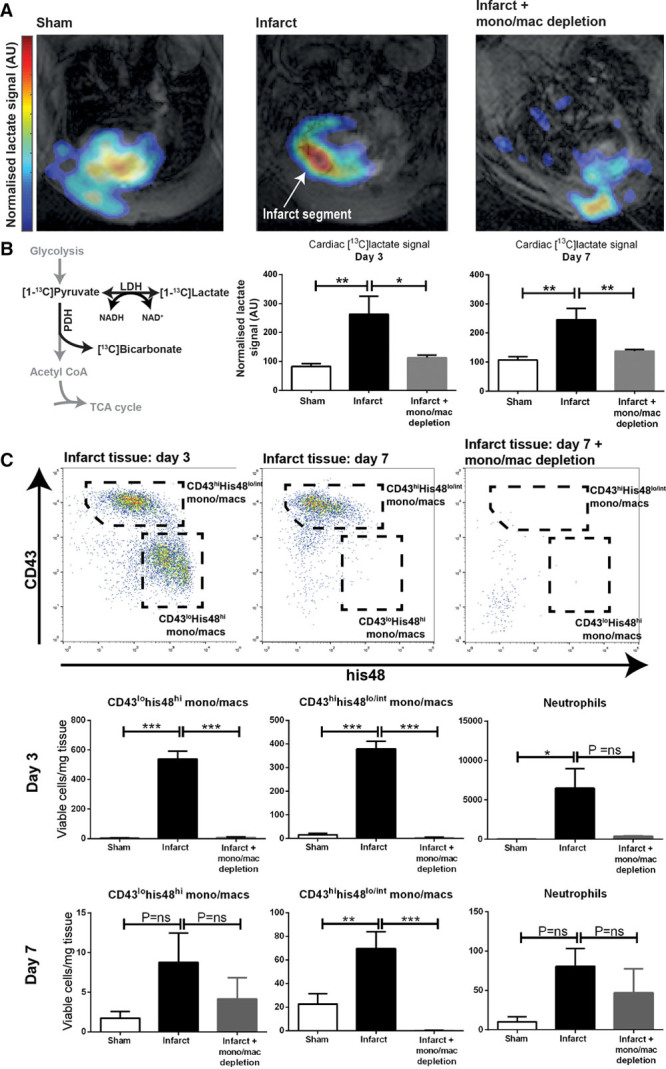
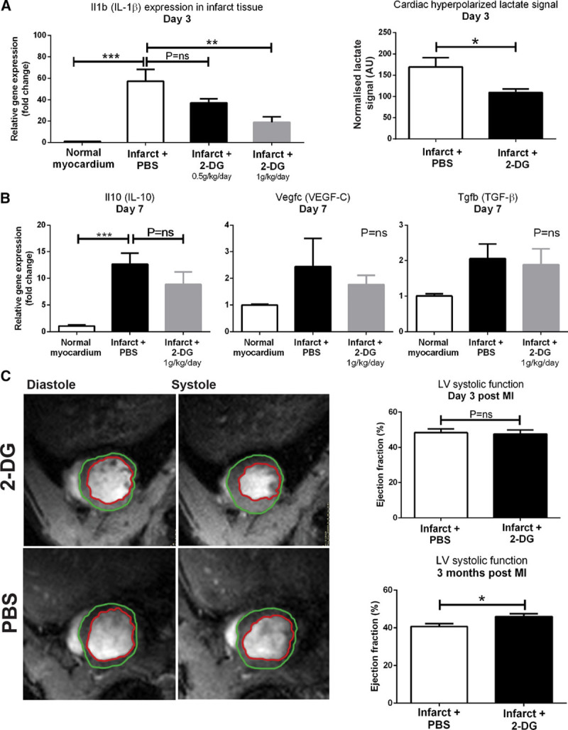
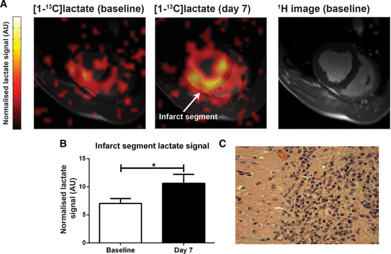
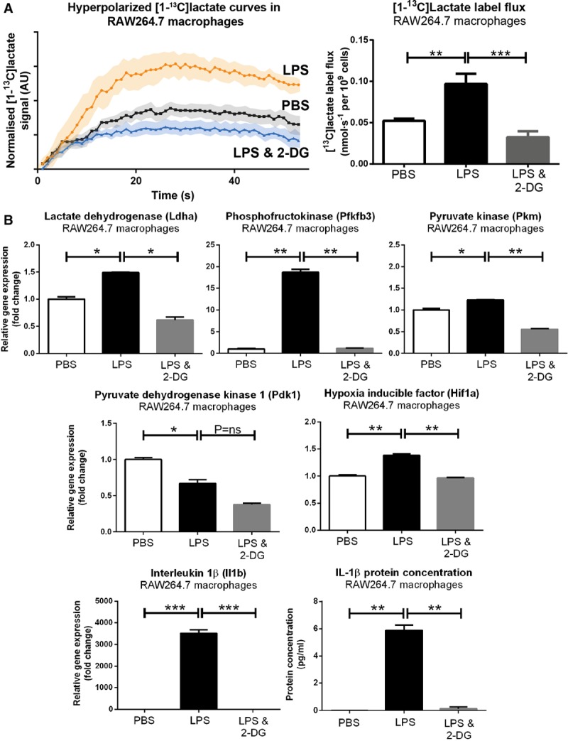
We performed hyperpolarized [1-C]pyruvate MR studies in small and large animal models of MI and in macrophage-like cell lines and measured the resulting [1-C]lactate signals. MI caused intense [1-C]lactate signal in healing myocardial segments at both day 3 and 7 after rodent MI, which was normalized at both time points after monocyte/macrophage depletion. A near-identical [1-C]lactate signature was also seen at day 7 after experimental MI in pigs. Hyperpolarized [1-C]pyruvate MR spectroscopy in macrophage-like cell suspensions demonstrated that macrophage activation and polarization with lipopolysaccharide almost doubled hyperpolarized lactate label flux rates in vitro; blockade of glycolysis with 2-deoxyglucose in activated cells normalized lactate label flux rates and markedly inhibited the production of key proinflammatory cytokines. Systemic administration of 2-deoxyglucose after rodent MI normalized the hyperpolarized [1-C]lactate signal in healing myocardial segments at day 3 and also caused dose-dependent improvement in IL (interleukin)-1β expression in infarct tissue without impairing the production of key reparative cytokines. Cine MRI demonstrated improvements in systolic function in 2-DG (2-deoxyglucose)-treated rats at 3 months.
Hyperpolarized MR using [1-C]pyruvate provides a novel method for the assessment of innate immune cell-driven inflammation in the heart after MI, with broad potential applicability across other cardiovascular disease states and suitability for early clinical translation.
目前的心血管临床成像技术仅提供对固有免疫细胞驱动的炎症的有限评估,固有免疫细胞驱动的炎症是心肌梗死 (MI) 和其他疾病的潜在治疗靶点。极化磁共振 (MR) 是一种新兴的成像技术,它可以生成对比度增强剂,使 MR 信号增强 10 到 20000 倍,从而实现心脏代谢物的映射。
确定使用 [1-C]丙酮酸的极化 MR 是否可以评估 MI 后局部心脏炎症反应。
我们在 MI 的小动物模型和巨噬细胞样细胞系中进行了极化 [1-C]丙酮酸的 MR 研究,并测量了由此产生的 [1-C]乳酸信号。MI 在啮齿动物 MI 后 3 天和 7 天,在愈合的心肌节段引起强烈的 [1-C]乳酸信号,在单核细胞/巨噬细胞耗竭后两个时间点均恢复正常。在猪的实验性 MI 后 7 天也观察到了几乎相同的 [1-C]乳酸特征。在巨噬细胞样细胞悬浮液中进行的极化 [1-C]丙酮酸磁共振波谱显示,用脂多糖激活和极化的巨噬细胞,体外极化的乳酸标签通量率几乎增加了一倍;用 2-脱氧葡萄糖阻断激活细胞中的糖酵解,使乳酸标签通量率正常化,并显著抑制关键前炎症细胞因子的产生。在 MI 后,在 MI 后 3 天,在愈合的心肌节段,用 2-脱氧葡萄糖进行全身给药,使极化 [1-C]乳酸信号正常化,也使梗死组织中白细胞介素 (IL)-1β的表达呈剂量依赖性改善,而不损害关键修复性细胞因子的产生。电影 MRI 显示,在 2-脱氧葡萄糖 (2-DG) 治疗的大鼠中,3 个月时收缩功能得到改善。
使用 [1-C]丙酮酸的极化磁共振提供了一种新的方法来评估 MI 后心脏中固有免疫细胞驱动的炎症,具有广泛的适用性,适用于其他心血管疾病状态,适合早期临床转化。