• 文献检索
  • 文档翻译
  • 深度研究
  • 学术资讯
  • Suppr Zotero 插件Zotero 插件
  • 邀请有礼
  • 套餐&价格
  • 历史记录
应用&插件
Suppr Zotero 插件Zotero 插件浏览器插件Mac 客户端Windows 客户端微信小程序
定价
高级版会员购买积分包购买API积分包
服务
文献检索文档翻译深度研究API 文档MCP 服务
关于我们
关于 Suppr公司介绍联系我们用户协议隐私条款
关注我们

Suppr 超能文献

核心技术专利:CN118964589B侵权必究
粤ICP备2023148730 号-1Suppr @ 2026

文献检索

告别复杂PubMed语法,用中文像聊天一样搜索,搜遍4000万医学文献。AI智能推荐,让科研检索更轻松。

立即免费搜索

文件翻译

保留排版,准确专业,支持PDF/Word/PPT等文件格式,支持 12+语言互译。

免费翻译文档

深度研究

AI帮你快速写综述,25分钟生成高质量综述,智能提取关键信息,辅助科研写作。

立即免费体验

一个大型的开源中风解剖大脑图像数据集和手动病变分割数据集。

A large, open source dataset of stroke anatomical brain images and manual lesion segmentations.

机构信息

University of Southern California, Los Angeles, California 90089, USA.

University of California, Irvine, Irvine, California 92697, USA.

出版信息

Sci Data. 2018 Feb 20;5:180011. doi: 10.1038/sdata.2018.11.

DOI:10.1038/sdata.2018.11
PMID:29461514
原文链接:https://pmc.ncbi.nlm.nih.gov/articles/PMC5819480/
Abstract

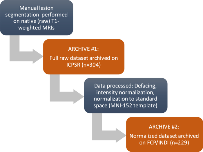
Stroke is the leading cause of adult disability worldwide, with up to two-thirds of individuals experiencing long-term disabilities. Large-scale neuroimaging studies have shown promise in identifying robust biomarkers (e.g., measures of brain structure) of long-term stroke recovery following rehabilitation. However, analyzing large rehabilitation-related datasets is problematic due to barriers in accurate stroke lesion segmentation. Manually-traced lesions are currently the gold standard for lesion segmentation on T1-weighted MRIs, but are labor intensive and require anatomical expertise. While algorithms have been developed to automate this process, the results often lack accuracy. Newer algorithms that employ machine-learning techniques are promising, yet these require large training datasets to optimize performance. Here we present ATLAS (Anatomical Tracings of Lesions After Stroke), an open-source dataset of 304 T1-weighted MRIs with manually segmented lesions and metadata. This large, diverse dataset can be used to train and test lesion segmentation algorithms and provides a standardized dataset for comparing the performance of different segmentation methods. We hope ATLAS release 1.1 will be a useful resource to assess and improve the accuracy of current lesion segmentation methods.

摘要

中风是全球导致成年人残疾的主要原因,多达三分之二的患者会长期残疾。大规模的神经影像学研究表明,在康复后识别长期中风恢复的强大生物标志物(例如大脑结构的测量值)方面具有前景。然而,由于在准确的中风损伤分割方面存在障碍,分析大规模的康复相关数据集是有问题的。手动追踪的损伤目前是 T1 加权 MRI 上损伤分割的金标准,但这项工作非常耗费人力,且需要具备解剖学专业知识。虽然已经开发了算法来自动完成此过程,但结果通常准确性不高。采用机器学习技术的较新算法很有前景,但这些算法需要大型训练数据集来优化性能。在这里,我们介绍了 ATLAS(中风后损伤的解剖追踪),这是一个包含 304 个 T1 加权 MRI 的开源数据集,具有手动分割的损伤和元数据。这个大型的、多样化的数据集可用于训练和测试损伤分割算法,并为比较不同分割方法的性能提供了一个标准化的数据集。我们希望 ATLAS 版本 1.1 将成为评估和提高当前损伤分割方法准确性的有用资源。

https://cdn.ncbi.nlm.nih.gov/pmc/blobs/fe83/5819480/aa34c526deec/sdata201811-f3.jpg
https://cdn.ncbi.nlm.nih.gov/pmc/blobs/fe83/5819480/7ded09f9360f/sdata201811-f1.jpg
https://cdn.ncbi.nlm.nih.gov/pmc/blobs/fe83/5819480/6db808ac7ea0/sdata201811-f2.jpg
https://cdn.ncbi.nlm.nih.gov/pmc/blobs/fe83/5819480/aa34c526deec/sdata201811-f3.jpg
https://cdn.ncbi.nlm.nih.gov/pmc/blobs/fe83/5819480/7ded09f9360f/sdata201811-f1.jpg
https://cdn.ncbi.nlm.nih.gov/pmc/blobs/fe83/5819480/6db808ac7ea0/sdata201811-f2.jpg
https://cdn.ncbi.nlm.nih.gov/pmc/blobs/fe83/5819480/aa34c526deec/sdata201811-f3.jpg

相似文献

1
A large, open source dataset of stroke anatomical brain images and manual lesion segmentations.一个大型的开源中风解剖大脑图像数据集和手动病变分割数据集。
Sci Data. 2018 Feb 20;5:180011. doi: 10.1038/sdata.2018.11.
2
A large, curated, open-source stroke neuroimaging dataset to improve lesion segmentation algorithms.一个大型、经过精心策划的、开源的中风神经影像学数据集,旨在改进病灶分割算法。
Sci Data. 2022 Jun 16;9(1):320. doi: 10.1038/s41597-022-01401-7.
3
Machine learning identifies stroke features between species.机器学习识别物种间的中风特征。
Theranostics. 2021 Jan 1;11(6):3017-3034. doi: 10.7150/thno.51887. eCollection 2021.
4
A comparison of automated lesion segmentation approaches for chronic stroke T1-weighted MRI data.慢性脑卒中风磁共振 T1 加权成像数据的自动病灶分割方法比较。
Hum Brain Mapp. 2019 Nov 1;40(16):4669-4685. doi: 10.1002/hbm.24729. Epub 2019 Jul 26.
5
Segmentation of stroke lesions using transformers-augmented MRI analysis.基于Transformer 增强 MRI 分析的脑卒中病灶分割。
Hum Brain Mapp. 2024 Aug 1;45(11):e26803. doi: 10.1002/hbm.26803.
6
Multi-atlas segmentation of the whole hippocampus and subfields using multiple automatically generated templates.使用多个自动生成的模板对整个海马体及其子区进行多图谱分割。
Neuroimage. 2014 Nov 1;101:494-512. doi: 10.1016/j.neuroimage.2014.04.054. Epub 2014 Apr 29.
7
Automatic Segmentation and Quantitative Assessment of Stroke Lesions on MR Images.磁共振图像上脑卒中标本的自动分割与定量评估
Diagnostics (Basel). 2022 Aug 24;12(9):2055. doi: 10.3390/diagnostics12092055.
8
An appraisal of the performance of AI tools for chronic stroke lesion segmentation.慢性脑卒中病灶分割人工智能工具性能评估。
Comput Biol Med. 2023 Sep;164:107302. doi: 10.1016/j.compbiomed.2023.107302. Epub 2023 Aug 1.
9
Lesion segmentation from multimodal MRI using random forest following ischemic stroke.基于随机森林的多模态 MRI 脑梗死病灶分割
Neuroimage. 2014 Sep;98:324-35. doi: 10.1016/j.neuroimage.2014.04.056. Epub 2014 May 2.
10
A large public dataset of annotated clinical MRIs and metadata of patients with acute stroke.一个大型的公共数据集,包含标注的临床 MRI 和急性中风患者的元数据。
Sci Data. 2023 Aug 22;10(1):548. doi: 10.1038/s41597-023-02457-9.

引用本文的文献

1
Functional Connectivity of Hippocampal Circuits and Visual Memory Function in Children and Adolescents With Perinatal Stroke.围产期卒中儿童和青少年海马回路的功能连接与视觉记忆功能
Hum Brain Mapp. 2025 Sep;46(13):e70342. doi: 10.1002/hbm.70342.
2
Unraveling Normal Anatomy via Fluid-Driven Anomaly Randomization.通过流体驱动的异常随机化解析正常解剖结构。
Proc IEEE Comput Soc Conf Comput Vis Pattern Recognit. 2025 Jun;2025:10455-10465. doi: 10.1109/cvpr52734.2025.00978. Epub 2025 Aug 13.
3
The future of data analysis is now: Integrating generative AI in neuroimaging methods development.

本文引用的文献

1
Heart Disease and Stroke Statistics-2017 Update: A Report From the American Heart Association.《2017年心脏病和中风统计数据更新:美国心脏协会报告》
Circulation. 2017 Mar 7;135(10):e146-e603. doi: 10.1161/CIR.0000000000000485. Epub 2017 Jan 25.
2
ISLES 2015 - A public evaluation benchmark for ischemic stroke lesion segmentation from multispectral MRI.ISLES 2015 - 多光谱 MRI 缺血性脑卒中病灶分割的公共评估基准。
Med Image Anal. 2017 Jan;35:250-269. doi: 10.1016/j.media.2016.07.009. Epub 2016 Jul 21.
3
Automated segmentation of chronic stroke lesions using LINDA: Lesion identification with neighborhood data analysis.
数据分析的未来已来:将生成式人工智能整合到神经成像方法开发中。
Imaging Neurosci (Camb). 2024 Jul 24;2. doi: 10.1162/imag_a_00241. eCollection 2024.
4
Enhanced nnU-Net Architectures for Automated MRI Segmentation of Head and Neck Tumors in Adaptive Radiation Therapy.用于自适应放射治疗中头颈部肿瘤自动MRI分割的增强型nnU-Net架构
Head Neck Tumor Segm MR Guid Appl (2024). 2025;15273:50-64. doi: 10.1007/978-3-031-83274-1_3. Epub 2025 Mar 3.
5
A concept-based interpretable model for the diagnosis of choroid neoplasias using multimodal data.一种基于概念的可解释模型,用于利用多模态数据诊断脉络膜肿瘤。
Nat Commun. 2025 Apr 13;16(1):3504. doi: 10.1038/s41467-025-58801-7.
6
FastSurfer-LIT: Lesion inpainting tool for whole-brain MRI segmentation with tumors, cavities, and abnormalities.FastSurfer-LIT:用于全脑MRI分割的病变修复工具,可处理肿瘤、空洞及异常情况。
Imaging Neurosci (Camb). 2025 Jan 31;3. doi: 10.1162/imag_a_00446. eCollection 2025 Jan 1.
7
Artificial intelligence in stroke risk assessment and management via retinal imaging.通过视网膜成像进行中风风险评估与管理的人工智能
Front Comput Neurosci. 2025 Feb 17;19:1490603. doi: 10.3389/fncom.2025.1490603. eCollection 2025.
8
Synthetic data in generalizable, learning-based neuroimaging.可推广的基于学习的神经影像学中的合成数据。
Imaging Neurosci (Camb). 2024 Nov 19;2:1-22. doi: 10.1162/imag_a_00337. eCollection 2024 Nov 1.
9
From MRIcro to MRIcron: The evolution of neuroimaging visualization tools.从MRIcro到MRIcron:神经影像可视化工具的演变。
Neuropsychologia. 2025 Jan 29;207:109067. doi: 10.1016/j.neuropsychologia.2025.109067. Epub 2025 Jan 4.
10
APIS: a paired CT-MRI dataset for ischemic stroke segmentation - methods and challenges.APIS:用于缺血性脑卒中分割的配对 CT-MRI 数据集——方法与挑战。
Sci Rep. 2024 Sep 4;14(1):20543. doi: 10.1038/s41598-024-71273-x.
使用LINDA对慢性中风病变进行自动分割:通过邻域数据分析进行病变识别。
Hum Brain Mapp. 2016 Apr;37(4):1405-21. doi: 10.1002/hbm.23110. Epub 2016 Jan 12.
4
Fast semi-automated lesion demarcation in stroke.中风中快速半自动病变划定
Neuroimage Clin. 2015 Jul 17;9:69-74. doi: 10.1016/j.nicl.2015.06.013. eCollection 2015.
5
Association between brain imaging signs, early and late outcomes, and response to intravenous alteplase after acute ischaemic stroke in the third International Stroke Trial (IST-3): secondary analysis of a randomised controlled trial.第三次国际卒中试验(IST-3)中急性缺血性卒中后脑成像征象、早期和晚期预后以及静脉注射阿替普酶反应之间的关联:一项随机对照试验的二次分析
Lancet Neurol. 2015 May;14(5):485-96. doi: 10.1016/S1474-4422(15)00012-5. Epub 2015 Mar 27.
6
Neural function, injury, and stroke subtype predict treatment gains after stroke.神经功能、损伤和中风亚型预测中风后的治疗效果。
Ann Neurol. 2015 Jan;77(1):132-45. doi: 10.1002/ana.24309. Epub 2014 Dec 4.
7
Global and regional burden of stroke during 1990-2010: findings from the Global Burden of Disease Study 2010.1990-2010 年全球及各区域卒中负担变化:来自 2010 年全球疾病负担研究的结果。
Lancet. 2014 Jan 18;383(9913):245-54. doi: 10.1016/s0140-6736(13)61953-4.
8
Apparent diffusion coefficient thresholds and diffusion lesion volume in acute stroke.急性脑卒中的表观扩散系数阈值和弥散病灶体积。
J Stroke Cerebrovasc Dis. 2013 Oct;22(7):906-9. doi: 10.1016/j.jstrokecerebrovasdis.2012.09.018. Epub 2012 Nov 24.
9
Early ischaemic diffusion lesion reduction in patients treated with intravenous tissue plasminogen activator: infrequent, but significantly associated with recanalization.静脉注射组织型纤溶酶原激活物治疗的患者早期缺血性弥散损伤减少:不常见,但与再通显著相关。
Int J Stroke. 2013 Jul;8(5):321-6. doi: 10.1111/j.1747-4949.2012.00902.x. Epub 2012 Sep 27.
10
Anatomy and physiology predict response to motor cortex stimulation after stroke.解剖学和生理学预测中风后运动皮层刺激的反应。
Neurology. 2011 Sep 13;77(11):1076-83. doi: 10.1212/WNL.0b013e31822e1482. Epub 2011 Aug 31.