Department of Computer Science and Engineering, Korea University, Seoul, Republic of Korea.
Department of Radiology, Kangbuk Samsung Medical Center, Seoul, Republic of Korea.
PLoS One. 2018 Sep 18;13(9):e0203355. doi: 10.1371/journal.pone.0203355. eCollection 2018.
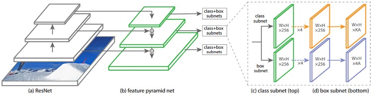
Several computer aided diagnosis (CAD) systems have been developed for mammography. They are widely used in certain countries such as the U.S. where mammography studies are conducted more frequently; however, they are not yet globally employed for clinical use due to their inconsistent performance, which can be attributed to their reliance on hand-crafted features. It is difficult to use hand-crafted features for mammogram images that vary due to factors such as the breast density of patients and differences in imaging devices. To address these problems, several studies have leveraged a deep convolutional neural network that does not require hand-crafted features. Among the recent object detectors, RetinaNet is particularly promising as it is a simpler one-stage object detector that is fast and efficient while achieving state-of-the-art performance. RetinaNet has been proven to perform conventional object detection tasks but has not been tested on detecting masses in mammograms. Thus, we propose a mass detection model based on RetinaNet. To validate its performance in diverse use cases, we construct several experimental setups using the public dataset INbreast and the in-house dataset GURO. In addition to training and testing on the same dataset (i.e., training and testing on INbreast), we evaluate our mass detection model in setups using additional training data (i.e., training on INbreast + GURO and testing on INbreast). We also evaluate our model in setups using pre-trained weights (i.e., using weights pre-trained on GURO, training and testing on INbreast). In all the experiments, our mass detection model achieves comparable or better performance than more complex state-of-the-art models including the two-stage object detector. Also, the results show that using the weights pre-trained on datasets achieves similar performance as directly using datasets in the training phase. Therefore, we make our mass detection model's weights pre-trained on both GURO and INbreast publicly available. We expect that researchers who train RetinaNet on their in-house dataset for the mass detection task can use our pre-trained weights to leverage the features extracted from the datasets.
已经开发了几种用于乳房 X 线照相术的计算机辅助诊断 (CAD) 系统。它们在美国等某些国家广泛使用,因为这些国家的乳房 X 线照相术检查更为频繁;但是,由于其性能不一致,这些系统尚未在全球范围内用于临床应用,这是因为它们依赖于手工制作的特征。对于因患者乳房密度和成像设备差异等因素而变化的乳房 X 线照片,使用手工制作的特征非常困难。为了解决这些问题,已经有几项研究利用了不需要手工制作特征的深度卷积神经网络。在最近的目标检测器中,RetinaNet 是一种特别有前途的单阶段目标检测器,它快速高效,同时实现了最先进的性能。已经证明 RetinaNet 可以执行常规的目标检测任务,但尚未在乳房 X 线照片中检测肿块。因此,我们提出了一种基于 RetinaNet 的肿块检测模型。为了验证其在不同用例中的性能,我们使用公共数据集 INbreast 和内部数据集 GURO 构建了几个实验设置。除了在同一数据集上进行训练和测试(即在 INbreast 上进行训练和测试)之外,我们还在使用其他训练数据的设置中评估了我们的肿块检测模型(即在 INbreast+GURO 上进行训练,在 INbreast 上进行测试)。我们还在使用预训练权重的设置中评估了我们的模型(即在 GURO 上预训练的权重,在 INbreast 上进行训练和测试)。在所有实验中,我们的肿块检测模型的性能都与包括两阶段目标检测器在内的更复杂的最先进模型相当或更好。此外,结果表明,使用在数据集上预训练的权重可以获得与在训练阶段直接使用数据集相同的性能。因此,我们公开了我们在 GURO 和 INbreast 上预训练的肿块检测模型的权重。我们希望那些在其内部数据集上训练用于肿块检测任务的 RetinaNet 的研究人员可以使用我们的预训练权重来利用从数据集提取的特征。