Rajeshkumar S
School of Bio-Sciences and Technology, VIT University, Vellore, TN, India.
J Genet Eng Biotechnol. 2016 Jun;14(1):195-202. doi: 10.1016/j.jgeb.2016.05.007. Epub 2016 Jun 9.
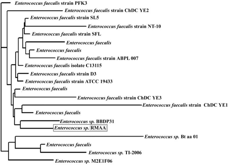
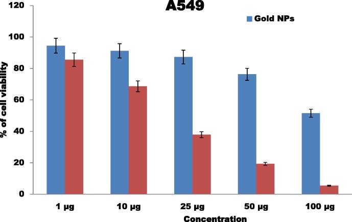
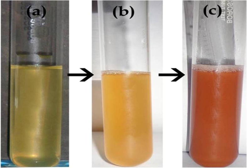
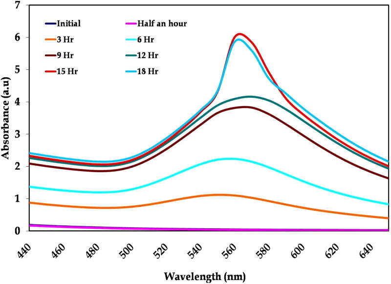
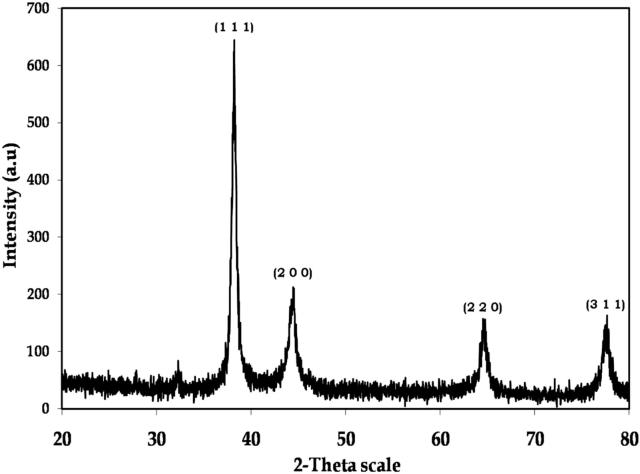
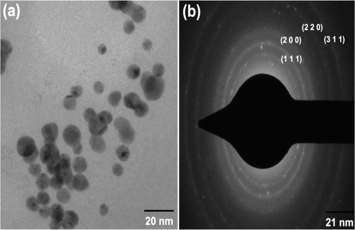
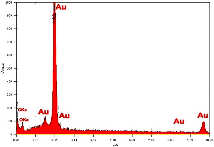
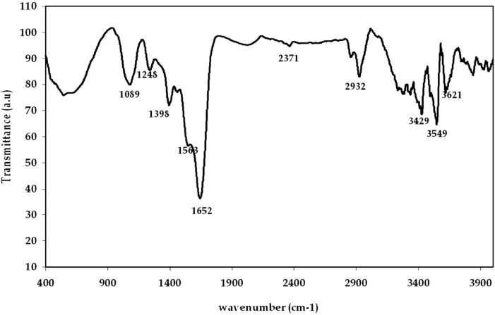
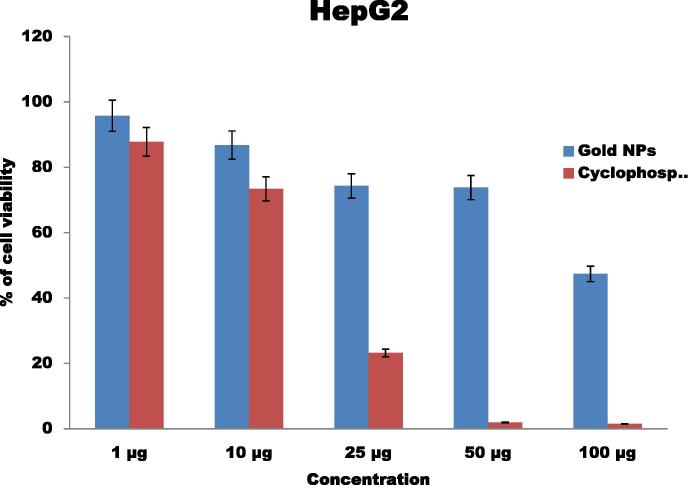
Gold nanoparticles have many applications in biomedical field. Improving delivery of anticancer agents to tumors using nanoparticles is one of the most promising research arenas in the field of nanotechnology. Eco-friendly gold nanoparticles synthesis was studied using marine bacteria sp. The nanoparticle synthesis started at 2 h of incubation time was identified by the formation of ruby red in the reaction mixture and SPR band centered at 545 nm. XRD shows that the strong four intense peaks indicate crystalline nature of nanoparticles. Morphology of nanoparticles analyzed by TEM shows that they are mostly spherical in shape with size ranging from 6 to 13 nm. EDX supports the presence of gold in the synthesized nanoparticles. FTIR reveals the active functional groups in the culture supernatant interaction with gold nanoparticles. As a result synthesized stable gold nanoparticles show more significant anticancer activity against HepG2 and A549 cells at 100 μg concentration of nanoparticles. This synthesis approach is simple, large scaled up a new door for development of targeted anticancer activity using gold nanoparticles and is novel in biomedical applications.
金纳米颗粒在生物医学领域有许多应用。利用纳米颗粒改善抗癌药物向肿瘤的递送是纳米技术领域最有前景的研究领域之一。使用海洋细菌菌株研究了环保型金纳米颗粒的合成。通过反应混合物中形成红宝石红色以及中心位于545nm的表面等离子体共振(SPR)带,确定在孵育2小时时开始合成纳米颗粒。X射线衍射(XRD)表明,四个强峰表明纳米颗粒具有晶体性质。通过透射电子显微镜(TEM)分析的纳米颗粒形态表明,它们大多为球形,尺寸范围为6至13nm。能量色散X射线光谱(EDX)证实合成的纳米颗粒中存在金。傅里叶变换红外光谱(FTIR)揭示了培养上清液中与金纳米颗粒相互作用的活性官能团。结果,在纳米颗粒浓度为100μg时,合成的稳定金纳米颗粒对肝癌细胞系(HepG2)和人肺癌细胞系(A549)显示出更显著的抗癌活性。这种合成方法简单,为利用金纳米颗粒开发靶向抗癌活性打开了一扇新的大门,并且在生物医学应用中是新颖的。