Department of Cardiology, The Second Affiliated Hospital of Wenzhou Medical University, 109 Xueyuan Road, Wenzhou 325027, Zhejiang, PR China.
Center for Molecular and Translational Medicine, Georgia State University, Research Science Center, 157 Decatur St SE, Atlanta, GA 30303. USA.
EBioMedicine. 2019 Mar;41:384-394. doi: 10.1016/j.ebiom.2019.02.032. Epub 2019 Feb 23.
FUN14 domain-containing 1 (FUNDC1), as a novel member of mitochondria-associated endoplasmic reticulum (ER) membranes associates with mitochondrial division and mitophagy. However, the expression profile and functional roles of FUNDC1 remain largely unclear in human cancer biology, including breast cancer (BC).
Immunohistochemistry and western blot analysis were used to determine the expression of FUNDC1 and BMI1 polycomb ring finger oncogene (BMI1). CCK8, cell counting and transwell assays were used to analyze cell proliferation, migration and invasion, respectively. Luciferase reporter and chromatin immunoprecipitation (ChIP) assays were used to detect the transcriptional regulation of Nuclear factor of activated T-cells, cytoplasmic 1 (NFATC1). The prognostic merit of NFATC1 expression was assessed by Kaplan-Meier assay.
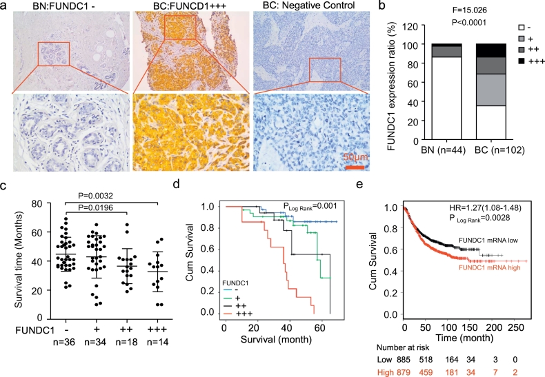
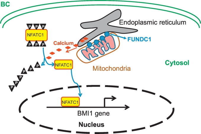
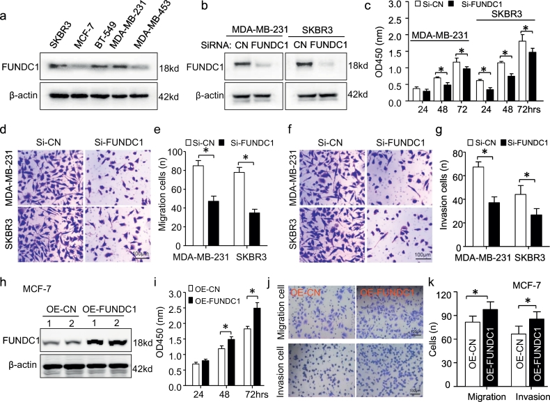
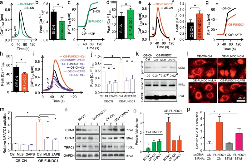
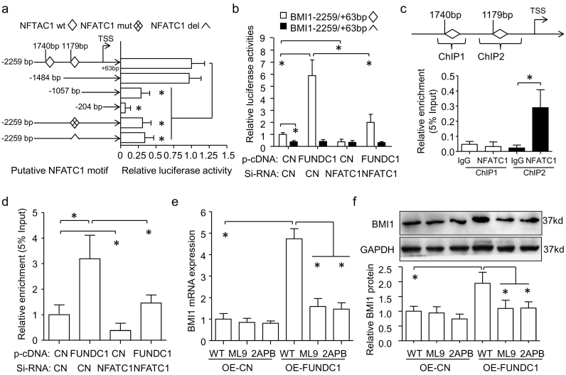
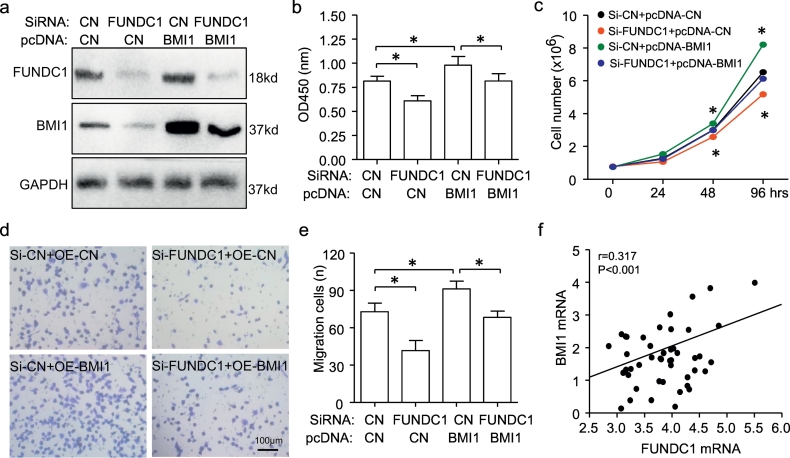
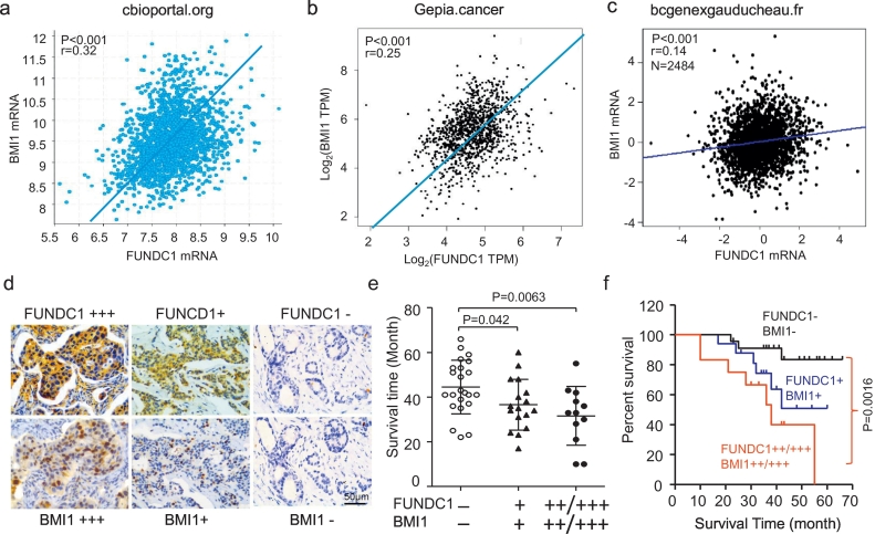
Immunohistochemistry revealed strong immunostaining for FUNDC1 in cytoplasmic and nuclear membrane distribution in BC tissues as compared with normal breast epithelium. Kaplan-Meier survival analysis showed worse outcome for BC patients with high FUNDC1 expression. In vitro assay of gain- and loss-of-function of FUNDC1 suggested that FUNDC1 could stimulate BC cell proliferation, migration and invasion. Furthermore, elevated FUNDC1 level promoted Ca cytosol influx from ER and extracellular, as well as NFATC1 nuclear translocation and activity. Nuclear NFATC1 bound to the BMI1 gene promoter and transcriptionally upregulated its expression. Notably, BMI1 overexpression could rescue the loss of function of FUNDC1. Co-expression of FUNDC1 and BMI1 in BC patients predicted worse prognosis than without either expression.
FUNDC1 might promote BC progression by activating the Ca-NFATC1-BMI1 axis. This pathway may be promising for developing multiple targets for BC therapy.
作为一种新型的线粒体相关内质网(ER)膜相关蛋白, FUN14 结构域包含蛋白 1(FUNDC1)与线粒体分裂和噬线粒体有关。然而,FUNDC1 在人类癌症生物学中的表达谱和功能作用在很大程度上仍不清楚,包括乳腺癌(BC)。
免疫组织化学和 Western blot 分析用于确定 FUNDC1 和多梳抑制因子 BMI1 基因 polycomb 环指癌基因(BMI1)的表达。CCK8、细胞计数和 Transwell 分析分别用于分析细胞增殖、迁移和侵袭。荧光素酶报告和染色质免疫沉淀(ChIP)分析用于检测激活的 T 细胞核因子,细胞质 1(NFATC1)的转录调控。通过 Kaplan-Meier 分析评估 NFATC1 表达的预后价值。
免疫组织化学显示,与正常乳腺上皮相比,BC 组织中 FUNDC1 在细胞质和核膜分布中呈强免疫染色。Kaplan-Meier 生存分析显示,FUNDC1 高表达的 BC 患者预后较差。FUNDC1 的增益和失活功能的体外测定表明,FUNDC1 可以刺激 BC 细胞增殖、迁移和侵袭。此外,升高的 FUNDC1 水平促进 ER 和细胞外 Ca 细胞质内流,以及 NFATC1 核转位和活性。核 NFATC1 与 BMI1 基因启动子结合并转录上调其表达。值得注意的是,BMI1 的过表达可以挽救 FUNDC1 失活的功能。BC 患者中 FUNDC1 和 BMI1 的共表达比没有任何表达预测预后更差。
FUNDC1 可能通过激活 Ca-NFATC1-BMI1 轴促进 BC 进展。该途径可能为开发 BC 治疗的多个靶点提供有希望的途径。