Aprupe Lilija, Litjens Geert, Brinker Titus J, van der Laak Jeroen, Grabe Niels
Hamamatsu Tissue Imaging and Analysis (TIGA) Center, BioQuant, Heidelberg University, Heidelberg, Germany.
Department of Medical Oncology, National Center for Tumor Diseases (NCT), University Hospital Heidelberg, Heidelberg, Germany.
PeerJ. 2019 Apr 10;7:e6335. doi: 10.7717/peerj.6335. eCollection 2019.
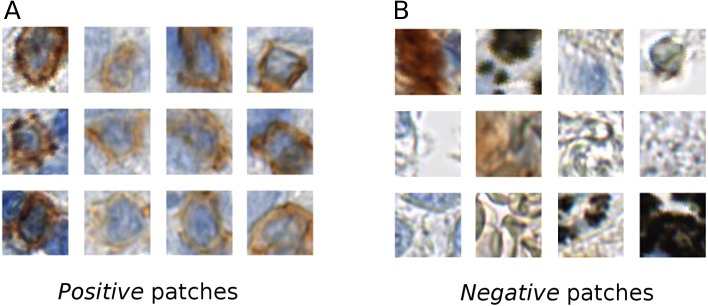
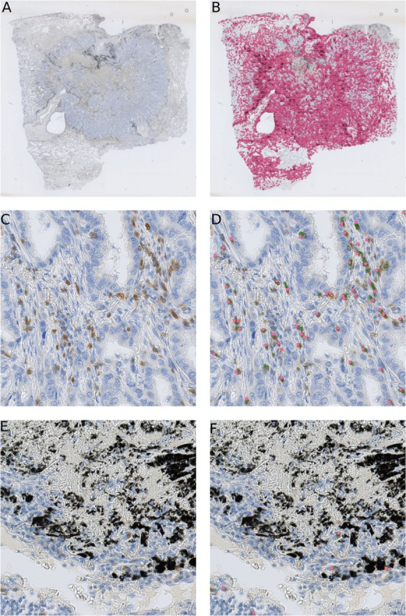
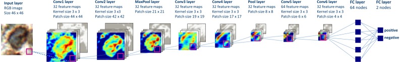
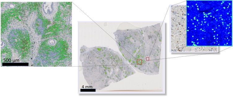
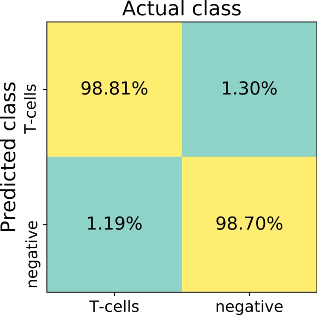
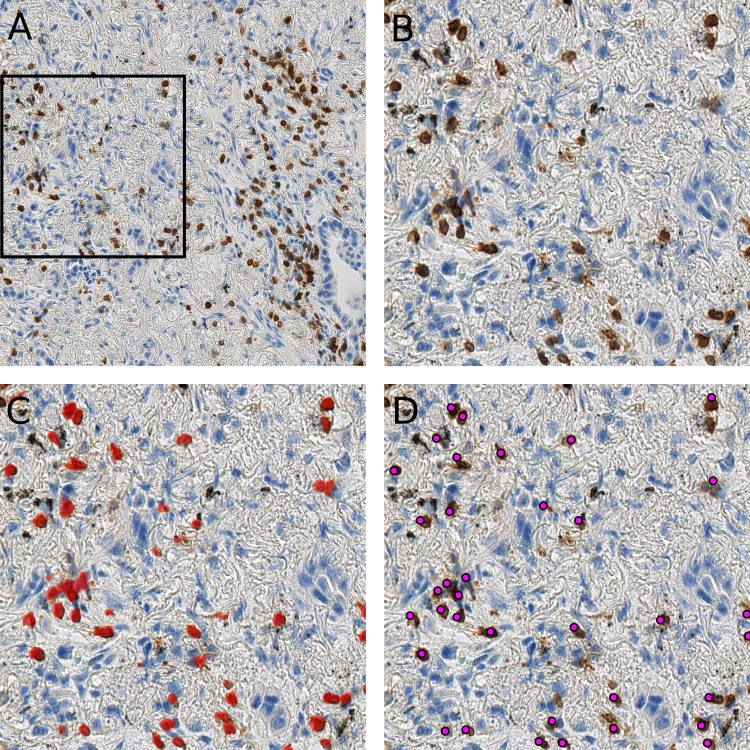
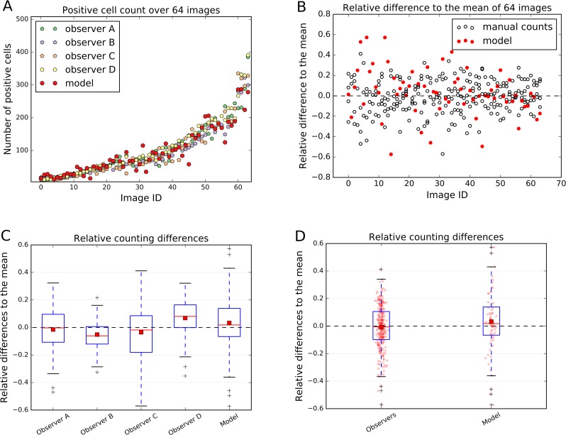
Recent years have seen a growing awareness of the role the immune system plays in successful cancer treatment, especially in novel therapies like immunotherapy. The characterization of the immunological composition of tumors and their micro-environment is thus becoming a necessity. In this paper we introduce a deep learning-based immune cell detection and quantification method, which is based on supervised learning, i.e., the input data for training comprises labeled images. Our approach objectively deals with staining variation and staining artifacts in immunohistochemically stained lung cancer tissue and is as precise as humans. This is evidenced by the low cell count difference to humans of 0.033 cells on average. This method, which is based on convolutional neural networks, has the potential to provide a new quantitative basis for research on immunotherapy.
近年来,人们越来越意识到免疫系统在成功的癌症治疗中所起的作用,尤其是在免疫疗法等新型疗法中。因此,对肿瘤及其微环境的免疫组成进行表征变得十分必要。在本文中,我们介绍了一种基于深度学习的免疫细胞检测和定量方法,该方法基于监督学习,即训练的输入数据包括带标记的图像。我们的方法客观地处理免疫组化染色肺癌组织中的染色变化和染色伪像,并且与人类一样精确。平均而言,与人类的细胞计数差异低至0.033个细胞,这证明了这一点。这种基于卷积神经网络的方法有可能为免疫疗法的研究提供新的定量基础。