Center for In Vivo Microscopy, Department of Radiology, Duke Medical Center, Duke University, 3302, Durham, NC, 27710, USA.
Department of Radiology, Duke University School of Medicine, Durham, NC, USA.
Brain Struct Funct. 2019 Jun;224(5):1797-1813. doi: 10.1007/s00429-019-01877-x. Epub 2019 Apr 20.
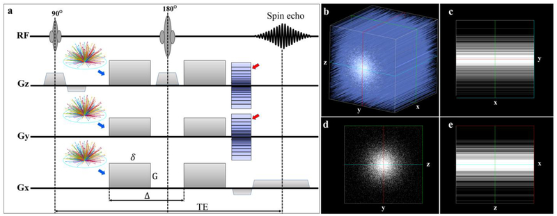
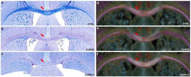
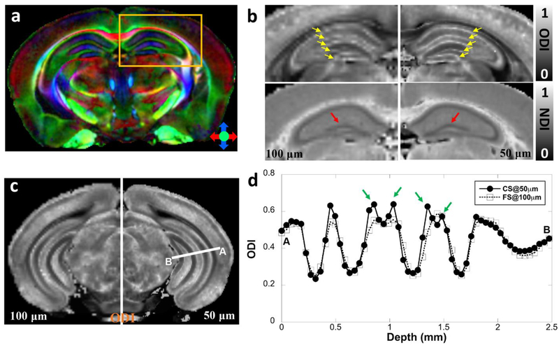
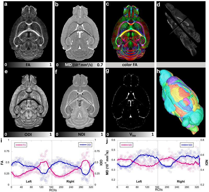
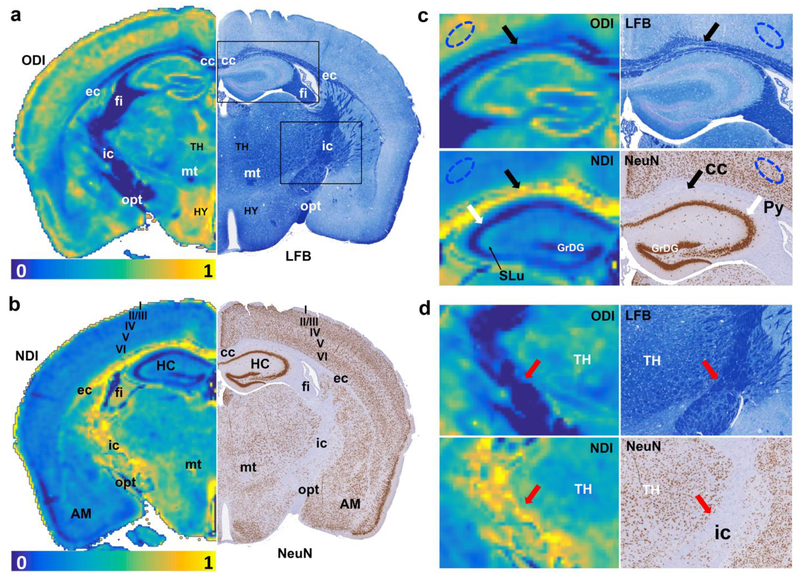
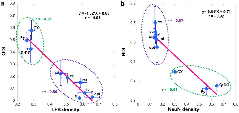
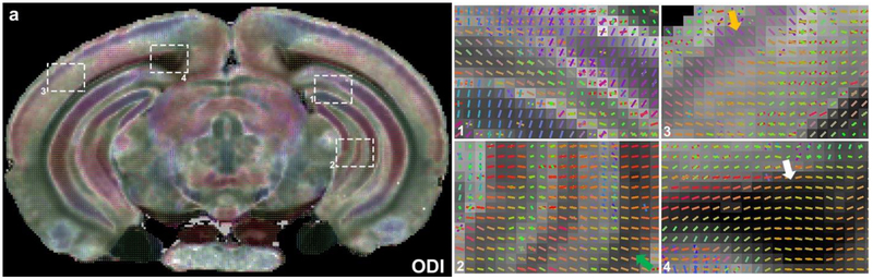
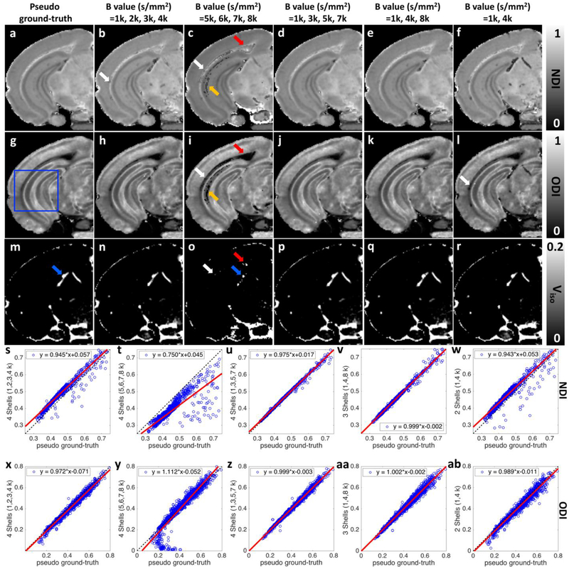
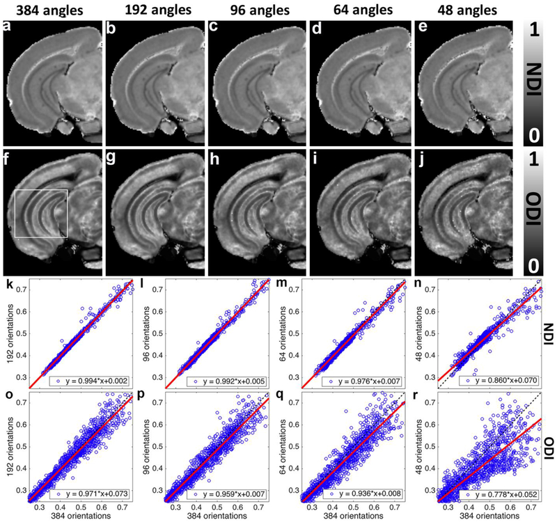
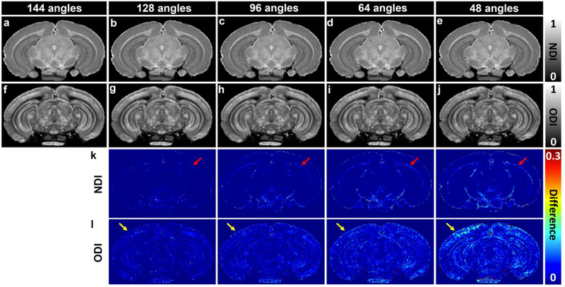
Advanced biophysical models like neurite orientation dispersion and density imaging (NODDI) have been developed to estimate the microstructural complexity of voxels enriched in dendrites and axons for both in vivo and ex vivo studies. NODDI metrics derived from high spatial and angular resolution diffusion MRI using the fixed mouse brain as a reference template have not yet been reported due in part to the extremely long scan time required. In this study, we modified the three-dimensional diffusion-weighted spin-echo pulse sequence for multi-shell and undersampling acquisition to reduce the scan time. This allowed us to acquire several exhaustive datasets that would otherwise not be attainable. NODDI metrics were derived from a complex 8-shell diffusion (1000-8000 s/mm) dataset with 384 diffusion gradient-encoding directions at 50 µm isotropic resolution. These provided a foundation for exploration of tradeoffs among acquisition parameters. A three-shell acquisition strategy covering low, medium, and high b values with at least angular resolution of 64 is essential for ex vivo NODDI experiments. The good agreement between neurite density index (NDI) and the orientation dispersion index (ODI) with the subsequent histochemical analysis of myelin and neuronal density highlights that NODDI could provide new insight into the microstructure of the brain. Furthermore, we found that NDI is sensitive to microstructural variations in the corpus callosum using a well-established demyelination cuprizone model. The study lays the ground work for developing protocols for routine use of high-resolution NODDI method in characterizing brain microstructure in mouse models.
高级生物物理模型,如神经突方向分散和密度成像(NODDI),已经被开发出来,以估计富含树突和轴突的体素的微观结构复杂性,用于体内和体外研究。由于需要非常长的扫描时间,因此尚未报道基于高空间和角度分辨率扩散 MRI 并使用固定小鼠大脑作为参考模板从 NODDI 指标导出的方法。在这项研究中,我们修改了用于多壳和欠采样采集的三维扩散加权自旋回波脉冲序列,以减少扫描时间。这使我们能够获取否则无法获得的几个详尽数据集。从具有 384 个扩散梯度编码方向的复杂 8 壳扩散(1000-8000 s/mm)数据集导出了 NODDI 指标,该数据集具有 50 µm 各向同性分辨率。这些为探索采集参数之间的权衡提供了基础。对于离体 NODDI 实验,具有至少 64 角分辨率的低、中、高 b 值的三壳采集策略对于获取离体 NODDI 实验至关重要。神经突密度指数(NDI)与取向分散指数(ODI)之间的良好一致性以及随后对髓磷脂和神经元密度的组织化学分析突出表明,NODDI 可以提供对大脑微观结构的新见解。此外,我们发现使用成熟的脱髓鞘杯状醇模型,NDI 对胼胝体的微观结构变化敏感。该研究为开发用于在小鼠模型中表征大脑微观结构的高分辨率 NODDI 方法的常规使用协议奠定了基础。