Martin-Garcia Jose M, Zhu Lan, Mendez Derek, Lee Ming-Yue, Chun Eugene, Li Chufeng, Hu Hao, Subramanian Ganesh, Kissick David, Ogata Craig, Henning Robert, Ishchenko Andrii, Dobson Zachary, Zhang Shangji, Weierstall Uwe, Spence John C H, Fromme Petra, Zatsepin Nadia A, Fischetti Robert F, Cherezov Vadim, Liu Wei
Biodesign Center for Applied Structural Discovery, Biodesign Institute, Arizona State University, 727 East Tyler Street, Tempe, AZ 85287, USA.
School of Molecular Sciences, Arizona State University, 551 East University Drive, Tempe, AZ 85287, USA.
IUCrJ. 2019 Apr 5;6(Pt 3):412-425. doi: 10.1107/S205225251900263X. eCollection 2019 May 1.
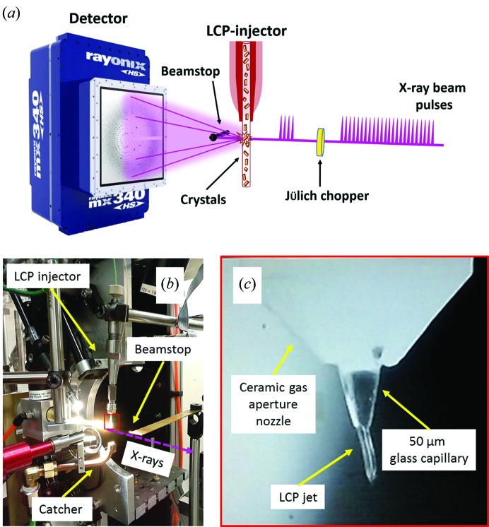
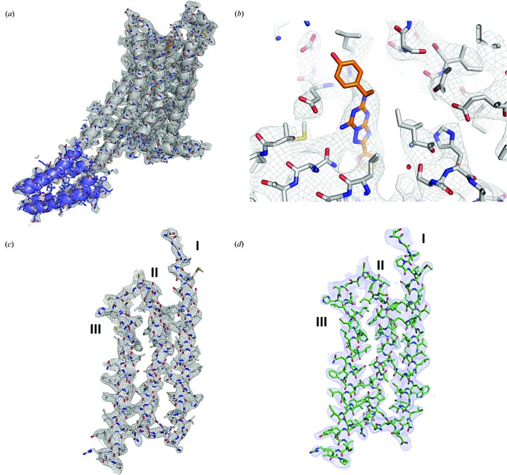
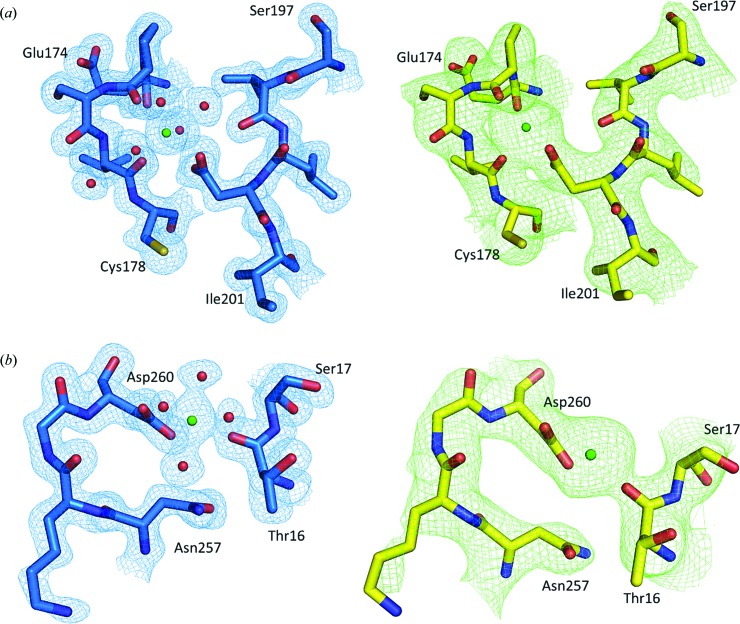
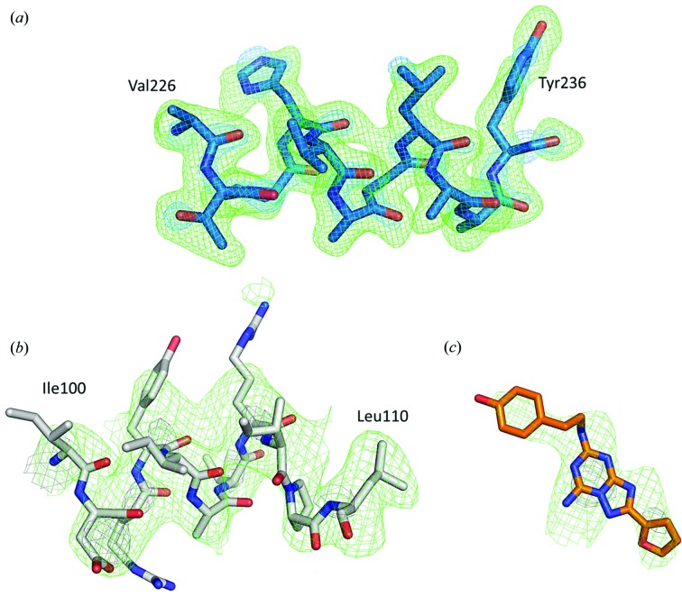
Since the first successful serial crystallography (SX) experiment at a synchrotron radiation source, the popularity of this approach has continued to grow showing that third-generation synchrotrons can be viable alternatives to scarce X-ray free-electron laser sources. Synchrotron radiation flux may be increased ∼100 times by a moderate increase in the bandwidth ('pink beam' conditions) at some cost to data analysis complexity. Here, we report the first high-viscosity injector-based pink-beam SX experiments. The structures of proteinase K (PK) and A adenosine receptor (AAR) were determined to resolutions of 1.8 and 4.2 Å using 4 and 24 consecutive 100 ps X-ray pulse exposures, respectively. Strong PK data were processed using existing Laue approaches, while weaker AAR data required an alternative data-processing strategy. This demonstration of the feasibility presents new opportunities for time-resolved experiments with microcrystals to study structural changes in real time at pink-beam synchrotron beamlines worldwide.
自从在同步辐射源上首次成功进行串行晶体学(SX)实验以来,这种方法的受欢迎程度持续增长,表明第三代同步加速器可以成为稀缺的X射线自由电子激光源的可行替代方案。通过适度增加带宽(“粉红光束”条件),同步辐射通量可增加约100倍,但会在一定程度上增加数据分析的复杂性。在此,我们报告了首次基于高粘度注射器的粉红光束SX实验。分别使用4次和24次连续的100 ps X射线脉冲曝光,将蛋白酶K(PK)和A腺苷受体(AAR)的结构解析到了1.8 Å和4.2 Å的分辨率。强PK数据使用现有的劳厄方法进行处理,而较弱的AAR数据则需要采用替代的数据处理策略。这种可行性的证明为在全球粉红光束同步加速器光束线上使用微晶进行时间分辨实验以实时研究结构变化提供了新机会。