Wang Duohao, Yao Qun, Yu Miao, Xiao Chaoyong, Fan Lin, Lin Xingjian, Zhu Donglin, Tian Minjie, Shi Jingping
Department of Neurology, Affiliated Brain Hospital of Nanjing Medical University, Nanjing, China.
Department of Radiology, Affiliated Brain Hospital of Nanjing Medical University, Nanjing, China.
Front Neurol. 2019 Jul 19;10:759. doi: 10.3389/fneur.2019.00759. eCollection 2019.
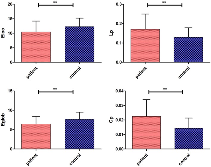
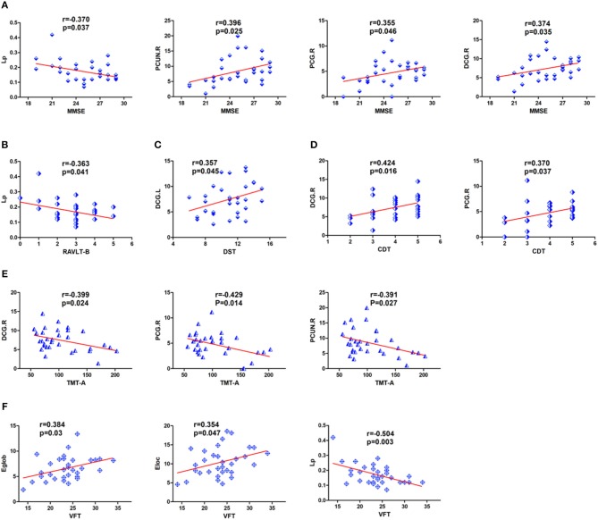
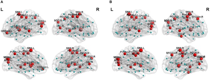
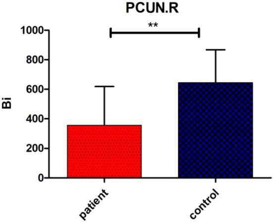
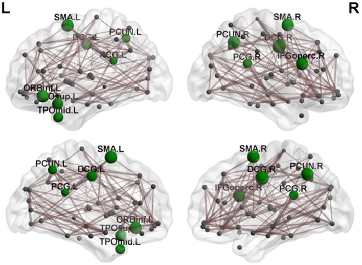
Cerebellar lesions can lead to a series of cognitive and emotional disorders by influencing cerebral activity via cerebro-cerebellar loops. To explore changes in cognitive function and structural brain networks in patients with posterior cerebellar infarction, we conducted the current study using diffusion-weighted MRI (32 cerebellar infarction patients, 29 controls). Moreover, a series of neuropsychological tests were used to assess the subject's cognitive performance. We found cognitive impairment following cerebellar infarction involving multiple cognitive domains, including memory, executive functions, visuospatial abilities, processing speed and language functions, and brain topological abnormalities, including changes in clustering coefficients, shortest path lengths, global efficiency, local efficiencies, betweenness centrality and nodal efficiencies. Our results indicated that measures of local efficiency, mainly in the precuneus, cingulate gyrus and frontal-temporal cortex, were significantly reduced with posterior cerebellar infarction. At the same time, The correlation analysis suggested thatthe abnormal alterations in the right PCG, bilateral DCG, right PCUN may play a core role in the cognitive impairment following cerebellar infarctions. The differences in topological features of the structural brain networks within the cerebro-cerebellar circuits may provide a new approach to explore the pathophysiological mechanisms of cognitive impairment following cerebellar infarction.
小脑病变可通过脑-小脑环路影响大脑活动,进而导致一系列认知和情感障碍。为探究小脑后梗死患者认知功能和脑结构网络的变化,我们使用扩散加权磁共振成像(32例小脑梗死患者,29例对照)进行了本研究。此外,还使用了一系列神经心理学测试来评估受试者的认知表现。我们发现小脑梗死后存在认知障碍,涉及多个认知领域,包括记忆、执行功能、视觉空间能力、处理速度和语言功能,以及脑拓扑异常,包括聚类系数、最短路径长度、全局效率、局部效率、介数中心性和节点效率的变化。我们的结果表明,主要在楔前叶、扣带回和额颞叶皮质的局部效率测量值在小脑后梗死后显著降低。同时,相关性分析表明,右侧顶下小叶、双侧扣带回、右侧楔前叶的异常改变可能在小脑梗死后的认知障碍中起核心作用。脑-小脑回路内结构脑网络拓扑特征的差异可能为探索小脑梗死后认知障碍的病理生理机制提供一种新方法。