Wanjare Maureen, Kawamura Masashi, Hu Caroline, Alcazar Cynthia, Wang Hanjay, Woo Y Joseph, Huang Ngan F
Center for Tissue Regeneration, Repair and Restoration, Veterans Affairs Palo Alto Health Care System, Palo Alto, CA, United States.
Stanford Cardiovascular Institute, Stanford University, Stanford, CA, United States.
Front Bioeng Biotechnol. 2019 Sep 3;7:208. doi: 10.3389/fbioe.2019.00208. eCollection 2019.
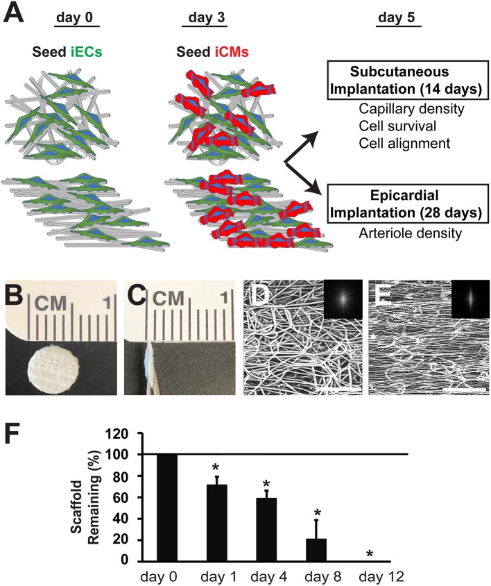
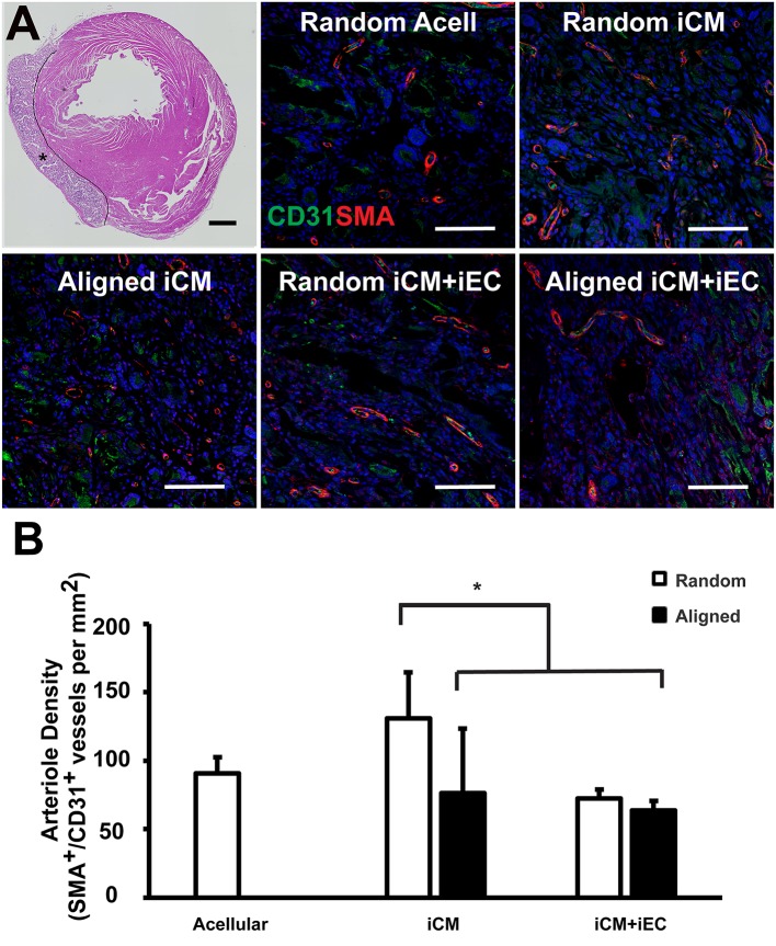
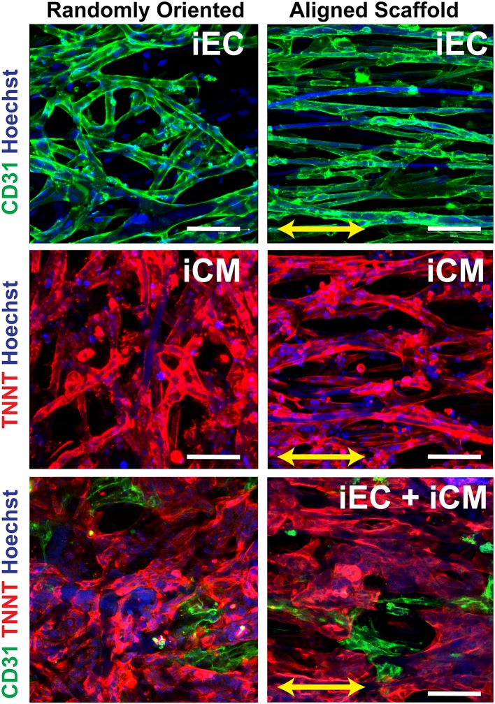
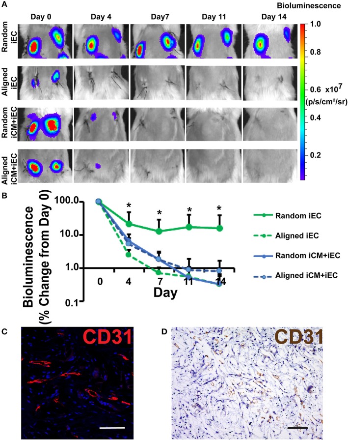
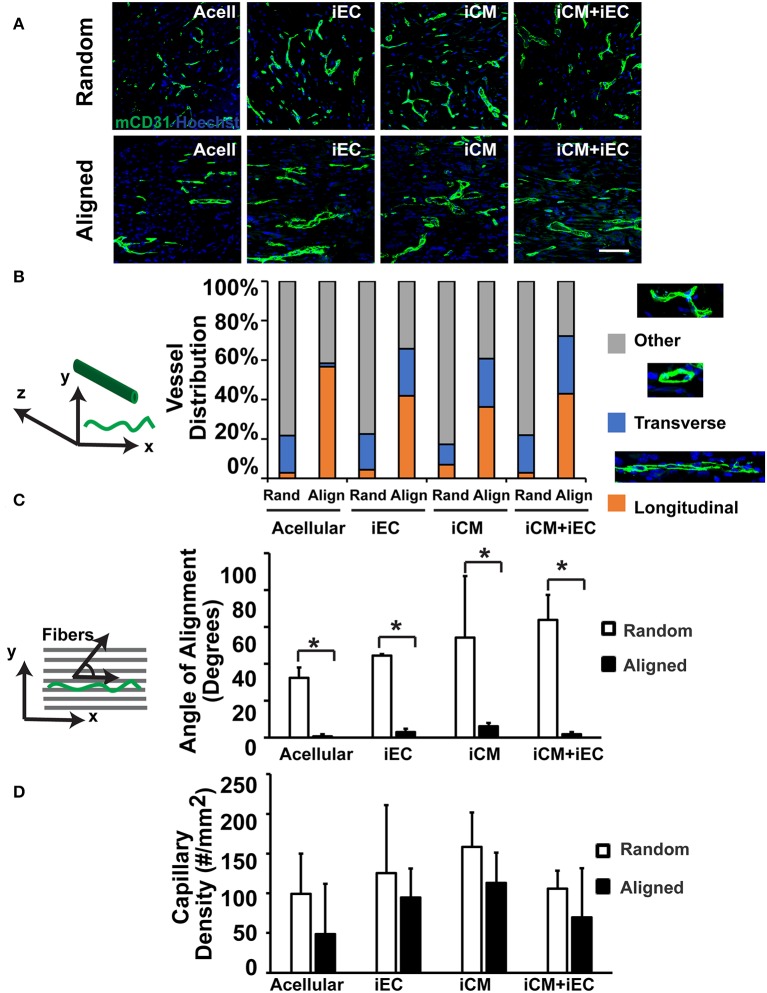
Tissue engineering approaches to regenerate myocardial tissue after disease or injury is promising. Integration with the host vasculature is critical to the survival and therapeutic efficacy of engineered myocardial tissues. To create more physiologically oriented engineered myocardial tissue with organized cellular arrangements and endothelial interactions, randomly oriented or parallel-aligned microfibrous polycaprolactone scaffolds were seeded with human pluripotent stem cell-derived cardiomyocytes (iCMs) and/or endothelial cells (iECs). The resultant engineered myocardial tissues were assessed in a subcutaneous transplantation model and in a myocardial injury model to evaluate the effect of scaffold anisotropy and endothelial interactions on vascular integration of the engineered myocardial tissue. Here we demonstrated that engineered myocardial tissue composed of randomly oriented scaffolds seeded with iECs promoted the survival of iECs for up to 14 days. However, engineered myocardial tissue composed of aligned scaffolds preferentially guided the organization of host capillaries along the direction of the microfibers. In a myocardial injury model, epicardially transplanted engineered myocardial tissues composed of randomly oriented scaffolds seeded with iCMs augmented microvessel formation leading to a significantly higher arteriole density after 4 weeks, compared to engineered tissues derived from aligned scaffolds. These findings that the scaffold microtopography imparts differential effect on revascularization, in which randomly oriented scaffolds promote pro-survival and pro-angiogenic effects, and aligned scaffolds direct the formation of anisotropic vessels. These findings suggest a dominant role of scaffold topography over endothelial co-culture in modulating cellular survival, vascularization, and microvessel architecture.
利用组织工程方法在疾病或损伤后再生心肌组织很有前景。与宿主脉管系统整合对于工程化心肌组织的存活和治疗效果至关重要。为了创建具有更生理导向性、细胞排列有序且有内皮细胞相互作用的工程化心肌组织,将人多能干细胞衍生的心肌细胞(iCMs)和/或内皮细胞(iECs)接种到随机取向或平行排列的微纤维聚己内酯支架上。在皮下移植模型和心肌损伤模型中对所得的工程化心肌组织进行评估,以评价支架各向异性和内皮细胞相互作用对工程化心肌组织血管整合的影响。在此我们证明,由接种了iECs的随机取向支架组成的工程化心肌组织可促进iECs存活长达14天。然而,由排列支架组成的工程化心肌组织优先引导宿主毛细血管沿微纤维方向排列。在心肌损伤模型中,与由排列支架衍生的工程化组织相比,经心外膜移植的、由接种了iCMs的随机取向支架组成的工程化心肌组织在4周后增加了微血管形成,导致小动脉密度显著更高。这些发现表明支架微观形貌对血管再生具有不同影响,其中随机取向支架促进细胞存活和促血管生成作用,而排列支架引导各向异性血管的形成。这些发现表明在调节细胞存活、血管化和微血管结构方面,支架形貌比内皮细胞共培养起更主要的作用。