Chen Yilong, Wang Kai, Liao Xiangyun, Qian Yinling, Wang Qiong, Yuan Zhiyong, Heng Pheng-Ann
Shenzhen Institutes of Advanced Technology, Chinese Academy of Sciences, Shenzhen, China.
AI Research Center, Peng Cheng Laboratory, Shenzhen, China.
Front Genet. 2019 Nov 26;10:1110. doi: 10.3389/fgene.2019.01110. eCollection 2019.
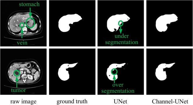
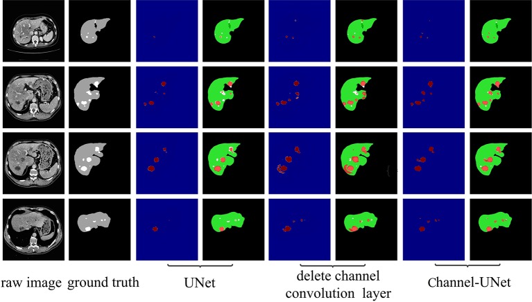
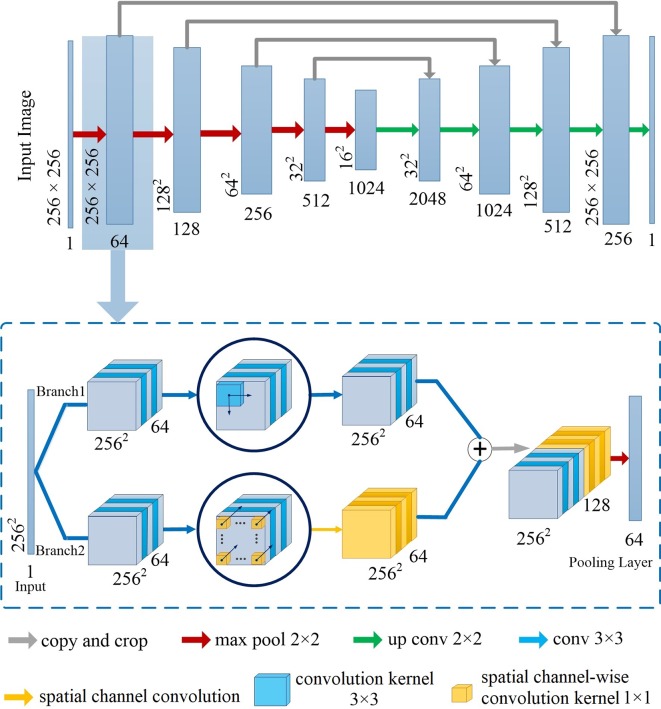
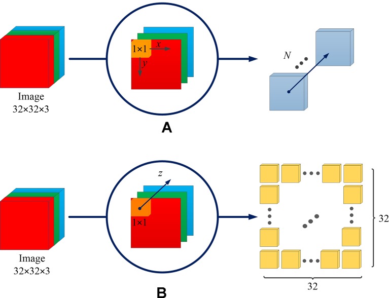
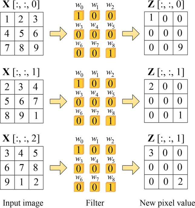
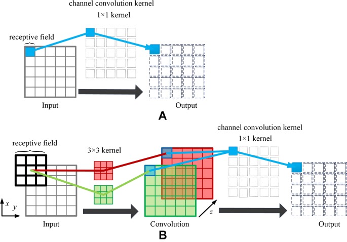
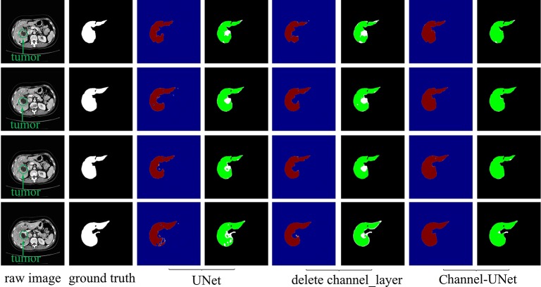
It is a challenge to automatically and accurately segment the liver and tumors in computed tomography (CT) images, as the problem of over-segmentation or under-segmentation often appears when the Hounsfield unit (Hu) of liver and tumors is close to the Hu of other tissues or background. In this paper, we propose the spatial channel-wise convolution, a convolutional operation along the direction of the channel of feature maps, to extract mapping relationship of spatial information between pixels, which facilitates learning the mapping relationship between pixels in the feature maps and distinguishing the tumors from the liver tissue. In addition, we put forward an iterative extending learning strategy, which optimizes the mapping relationship of spatial information between pixels at different scales and enables spatial channel-wise convolution to map the spatial information between pixels in high-level feature maps. Finally, we propose an end-to-end convolutional neural network called Channel-UNet, which takes UNet as the main structure of the network and adds spatial channel-wise convolution in each up-sampling and down-sampling module. The network can converge the optimized mapping relationship of spatial information between pixels extracted by spatial channel-wise convolution and information extracted by feature maps and realizes multi-scale information fusion. The proposed ChannelUNet is validated by the segmentation task on the 3Dircadb dataset. The Dice values of liver and tumors segmentation were 0.984 and 0.940, which is slightly superior to current best performance. Besides, compared with the current best method, the number of parameters of our method reduces by 25.7%, and the training time of our method reduces by 33.3%. The experimental results demonstrate the efficiency and high accuracy of Channel-UNet in liver and tumors segmentation in CT images.
在计算机断层扫描(CT)图像中自动且准确地分割肝脏和肿瘤是一项挑战,因为当肝脏和肿瘤的亨氏单位(Hu)与其他组织或背景的Hu接近时,常常会出现过分割或欠分割的问题。在本文中,我们提出了空间通道卷积,这是一种沿着特征图通道方向的卷积操作,用于提取像素之间空间信息的映射关系,这有助于学习特征图中像素之间的映射关系,并将肿瘤与肝脏组织区分开来。此外,我们提出了一种迭代扩展学习策略,该策略优化了不同尺度下像素之间空间信息的映射关系,并使空间通道卷积能够映射高级特征图中像素之间的空间信息。最后,我们提出了一种名为Channel-UNet的端到端卷积神经网络,它以UNet作为网络的主要结构,并在每个上采样和下采样模块中添加空间通道卷积。该网络可以融合由空间通道卷积提取的像素之间空间信息的优化映射关系和由特征图提取的信息,并实现多尺度信息融合。所提出的ChannelUNet在3Dircadb数据集上的分割任务中得到了验证。肝脏和肿瘤分割的Dice值分别为0.984和0.940,略优于当前的最佳性能。此外,与当前最佳方法相比,我们方法的参数数量减少了25.7%,训练时间减少了33.3%。实验结果证明了Channel-UNet在CT图像中肝脏和肿瘤分割方面的效率和高精度。