Department of Gastroenterology, Sir Run Run Shaw Hospital, Medical School, Zhejiang University, Hangzhou, China.
Institute of Gastroenterology, Zhejiang University (IGZJU), Hangzhou, China.
Clin Transl Gastroenterol. 2019 Dec;10(12):e00109. doi: 10.14309/ctg.0000000000000109.
Application of artificial intelligence in gastrointestinal endoscopy is increasing. The aim of the study was to examine the accuracy of convolutional neural network (CNN) using endoscopic images for evaluating Helicobacter pylori (H. pylori) infection.
Patients who received upper endoscopy and gastric biopsies at Sir Run Run Shaw Hospital (January 2015-June 2015) were retrospectively searched. A novel Computer-Aided Decision Support System that incorporates CNN model (ResNet-50) based on endoscopic gastric images was developed to evaluate for H. pylori infection. Diagnostic accuracy was evaluated in an independent validation cohort. H. pylori infection was defined by the presence of H. pylori on immunohistochemistry testing on gastric biopsies and/or a positive 13C-urea breath test.
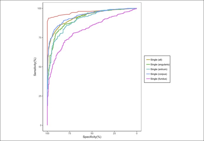
Of 1,959 patients, 1,507 (77%) including 847 (56%) with H. pylori infection (11,729 gastric images) were assigned to the derivation cohort, and 452 (23%) including 310 (69%) with H. pylori infection (3,755 images) were assigned to the validation cohort. The area under the curve for a single gastric image was 0.93 (95% confidence interval [CI] 0.92-0.94) with sensitivity, specificity, and accuracy of 81.4% (95% CI 79.8%-82.9%), 90.1% (95% CI 88.4%-91.7%), and 84.5% (95% CI 83.3%-85.7%), respectively, using an optimal cutoff value of 0.3. Area under the curve for multiple gastric images (8.3 ± 3.3) per patient was 0.97 (95% CI 0.96-0.99) with sensitivity, specificity, and accuracy of 91.6% (95% CI 88.0%-94.4%), 98.6% (95% CI 95.0%-99.8%), and 93.8% (95% CI 91.2%-95.8%), respectively, using an optimal cutoff value of 0.4.
In this pilot study, CNN using multiple archived gastric images achieved high diagnostic accuracy for the evaluation of H. pylori infection.
人工智能在胃肠内镜中的应用正在增加。本研究旨在通过内镜图像检查卷积神经网络(CNN)评估幽门螺杆菌(H. pylori)感染的准确性。
回顾性检索了 2015 年 1 月至 2015 年 6 月在浙江大学医学院附属邵逸夫医院接受上消化道内镜检查和胃活检的患者。开发了一种新的计算机辅助决策支持系统,该系统基于内镜胃图像纳入 CNN 模型(ResNet-50),用于评估 H. pylori 感染。在独立验证队列中评估诊断准确性。H. pylori 感染通过胃活检免疫组织化学检测 H. pylori 阳性和/或 13C-尿素呼气试验阳性来定义。
在 1959 例患者中,1507 例(77%)包括 847 例(56%)H. pylori 感染患者(11729 张胃图像)被分配到推导队列,452 例(23%)包括 310 例(69%)H. pylori 感染患者(3755 张图像)被分配到验证队列。单个胃图像的曲线下面积为 0.93(95%置信区间 [CI] 0.92-0.94),其灵敏度、特异性和准确率分别为 81.4%(95% CI 79.8%-82.9%)、90.1%(95% CI 88.4%-91.7%)和 84.5%(95% CI 83.3%-85.7%),最佳截断值为 0.3。每位患者的多个胃图像(8.3±3.3)的曲线下面积为 0.97(95% CI 0.96-0.99),灵敏度、特异性和准确率分别为 91.6%(95% CI 88.0%-94.4%)、98.6%(95% CI 95.0%-99.8%)和 93.8%(95% CI 91.2%-95.8%),最佳截断值为 0.4。
在这项初步研究中,使用多个存档胃图像的 CNN 实现了 H. pylori 感染评估的高诊断准确性。