IEEE Trans Ultrason Ferroelectr Freq Control. 2021 Apr;68(4):1096-1104. doi: 10.1109/TUFFC.2020.3033304. Epub 2021 Mar 26.
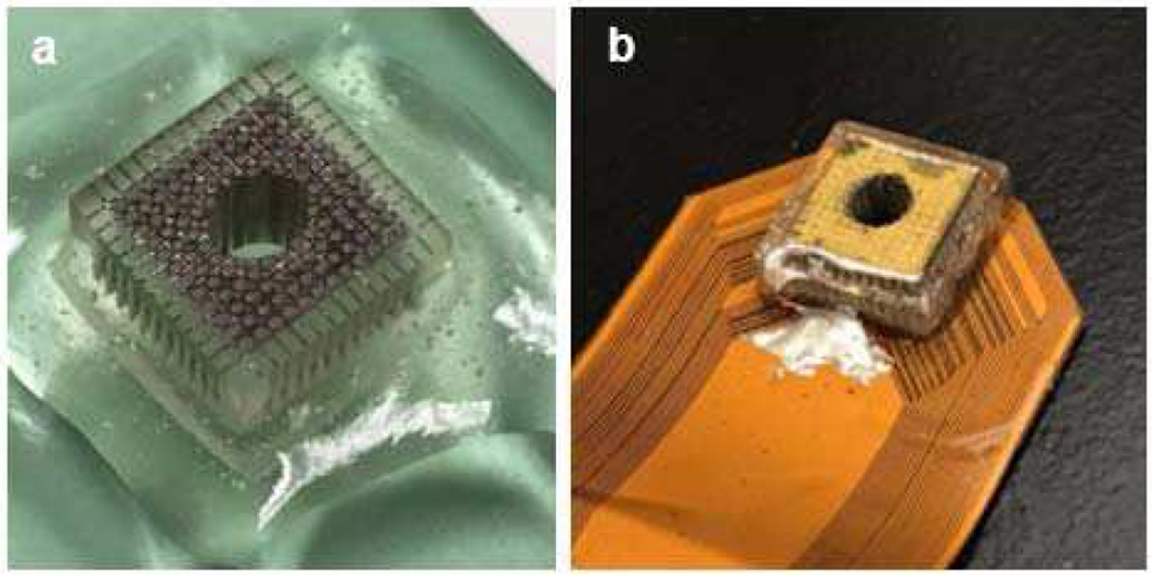
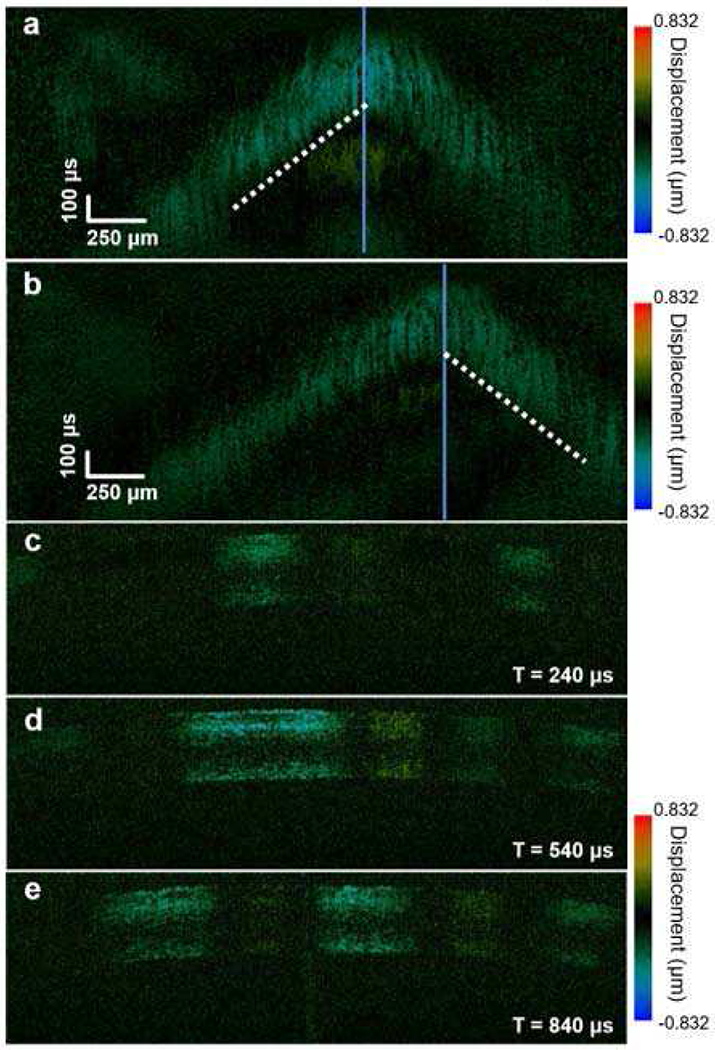
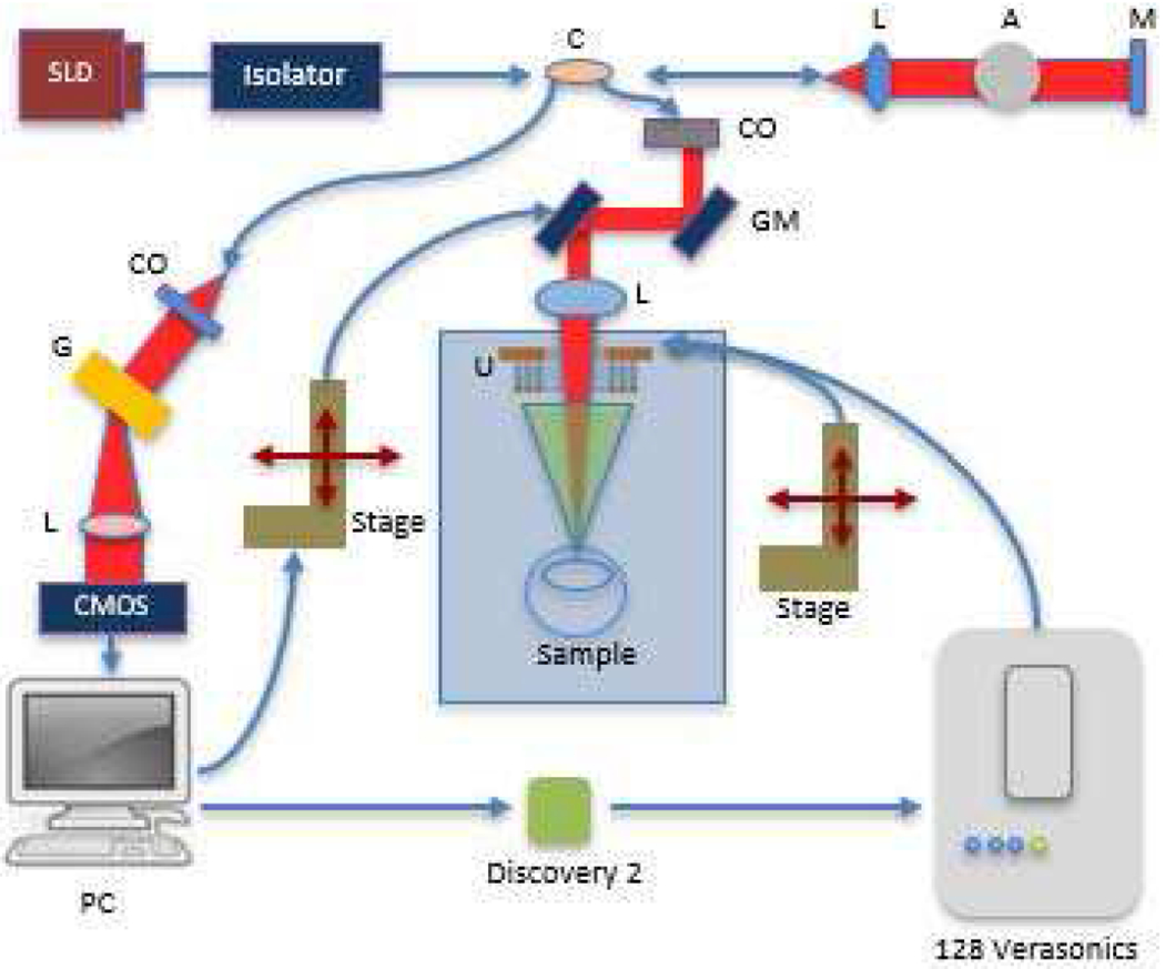
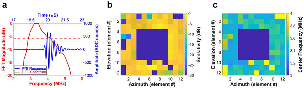
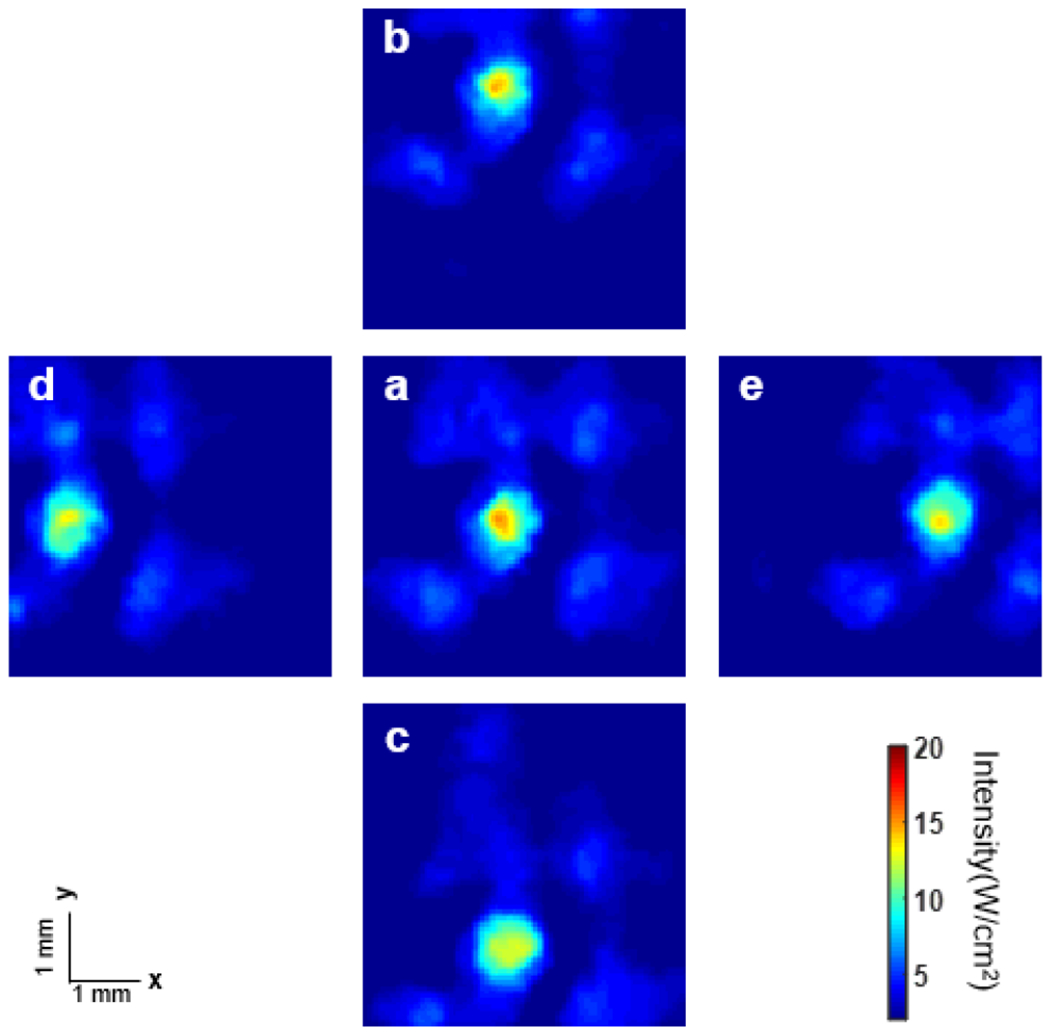
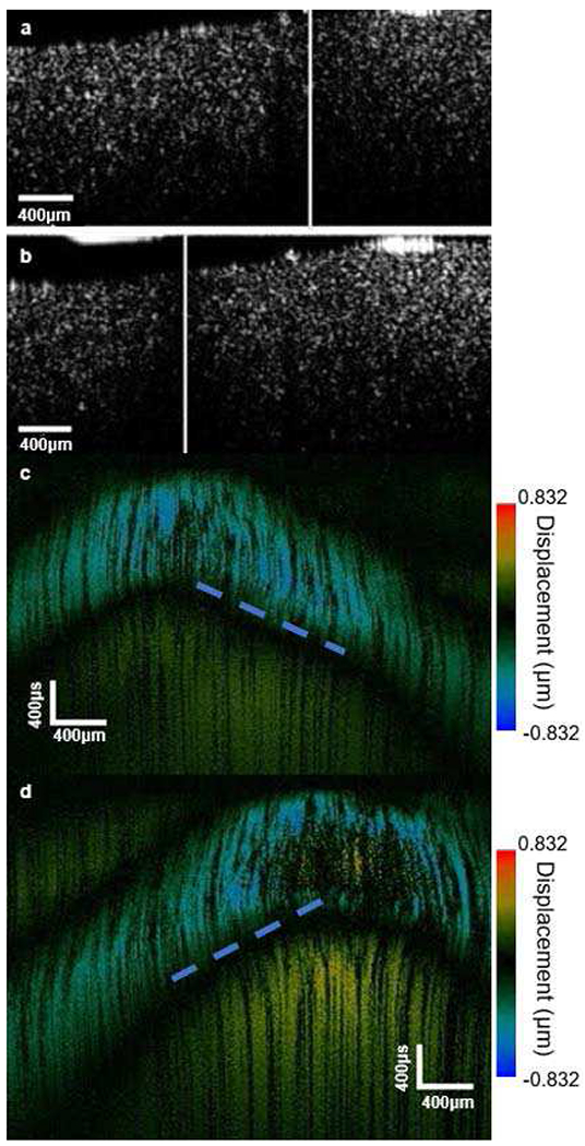
Acoustic radiation force optical coherence elastography (ARF-OCE) has been successfully implemented to characterize the biomechanical properties of soft tissues, such as the cornea and the retina, with high resolution using single-element ultrasonic transducers for ARF excitation. Most currently proposed OCE techniques, such as air puff and ARF, have less capability to control the spatiotemporal information of the induced region of deformation, resulting in limited accuracy and low temporal resolution of the shear wave elasticity imaging. In this study, we propose a new method called 2-D ultrasonic array-based OCE imaging, which combines the advantages of 3-D dynamic electronic steering of the 2-D ultrasonic array and high-resolution optical coherence tomography (OCT). The 3-D steering capability of the 2-D array was first validated using a hydrophone. Then, the combined 2-D ultrasonic array OCE system was calibrated using a homogenous phantom, followed by an experiment on ex vivo rabbit corneal tissue. The results demonstrate that our newly developed 2-D ultrasonic array-based OCE system has the capability to map tissue biomechanical properties accurately, and therefore, has the potential to be a vital diagnostic tool in ophthalmology.
声辐射力光相干弹性成像(ARF-OCE)已成功应用于使用单个超声换能器进行声辐射力激发,以高分辨率对角膜和视网膜等软组织的生物力学特性进行特征描述。目前大多数提出的 OCE 技术,如空气脉冲和 ARF,对所产生的变形区域的时空信息的控制能力较弱,导致剪切波弹性成像的准确性有限和时间分辨率低。在这项研究中,我们提出了一种称为基于二维超声阵列的 OCE 成像的新方法,它结合了二维超声阵列的 3D 动态电子转向和高分辨率光学相干断层扫描(OCT)的优势。首先使用水听器验证了二维阵列的 3D 转向能力。然后,使用均匀的体模对组合的二维超声阵列 OCE 系统进行校准,随后对离体兔角膜组织进行实验。结果表明,我们新开发的基于二维超声阵列的 OCE 系统具有准确绘制组织生物力学特性的能力,因此有可能成为眼科诊断的重要工具。