J. Philip Kistler Stroke Research Center, Department of Neurology, Massachusetts General Hospital, Harvard Medical School, Boston, USA; Biomedical Imaging Group Rotterdam, Department of Radiology and Nuclear Medicine, Erasmus MC - University Medical Center Rotterdam, The Netherlands.
Biomedical Imaging Group Rotterdam, Department of Radiology and Nuclear Medicine, Erasmus MC - University Medical Center Rotterdam, The Netherlands; Department of Computer Science, University of Copenhagen, Copenhagen, Denmark.
Med Image Anal. 2020 Jul;63:101698. doi: 10.1016/j.media.2020.101698. Epub 2020 Apr 18.
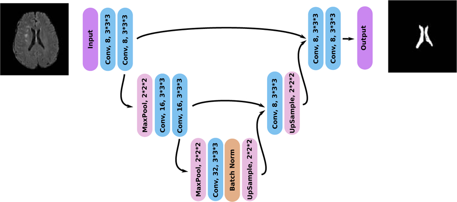
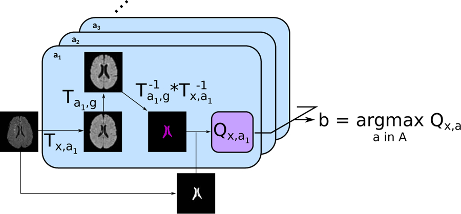
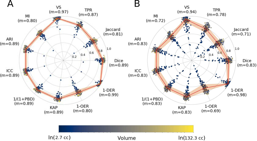
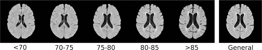
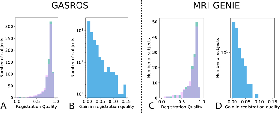
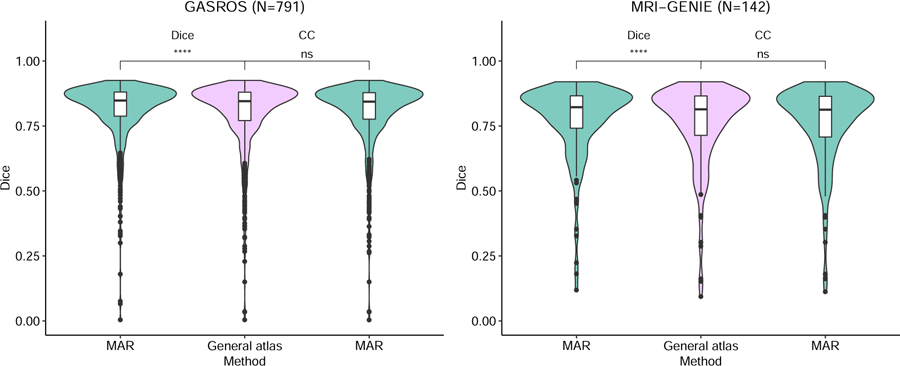
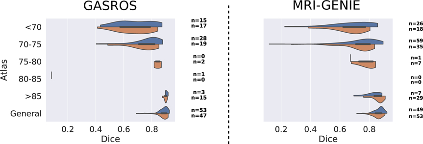
Registration is a core component of many imaging pipelines. In case of clinical scans, with lower resolution and sometimes substantial motion artifacts, registration can produce poor results. Visual assessment of registration quality in large clinical datasets is inefficient. In this work, we propose to automatically assess the quality of registration to an atlas in clinical FLAIR MRI scans of the brain. The method consists of automatically segmenting the ventricles of a given scan using a neural network, and comparing the segmentation to the atlas ventricles propagated to image space. We used the proposed method to improve clinical image registration to a general atlas by computing multiple registrations - one directly to the general atlas and others via different age-specific atlases - and then selecting the registration that yielded the highest ventricle overlap. Finally, as an example application of the complete pipeline, a voxelwise map of white matter hyperintensity burden was computed using only the scans with registration quality above a predefined threshold. Methods were evaluated in a single-site dataset of more than 1000 scans, as well as a multi-center dataset comprising 142 clinical scans from 12 sites. The automated ventricle segmentation reached a Dice coefficient with manual annotations of 0.89 in the single-site dataset, and 0.83 in the multi-center dataset. Registration via age-specific atlases could improve ventricle overlap compared to a direct registration to the general atlas (Dice similarity coefficient increase up to 0.15). Experiments also showed that selecting scans with the registration quality assessment method could improve the quality of average maps of white matter hyperintensity burden, instead of using all scans for the computation of the white matter hyperintensity map. In this work, we demonstrated the utility of an automated tool for assessing image registration quality in clinical scans. This image quality assessment step could ultimately assist in the translation of automated neuroimaging pipelines to the clinic.
注册是许多成像流水线的核心组成部分。在临床扫描的情况下,由于分辨率较低且有时存在大量运动伪影,因此注册可能会产生较差的结果。在大型临床数据集上,对注册质量进行视觉评估效率低下。在这项工作中,我们提出了一种自动评估临床 FLAIR MRI 脑扫描到图谱的配准质量的方法。该方法包括使用神经网络自动分割给定扫描的脑室,并将分割与传播到图像空间的图谱脑室进行比较。我们使用提出的方法通过计算多个配准(一个直接到通用图谱,另一个通过不同年龄特定的图谱)来改进到通用图谱的临床图像配准,然后选择产生最高脑室重叠的配准。最后,作为完整流水线的一个示例应用,仅使用配准质量超过预定义阈值的扫描计算了白质高信号负担的体素图。方法在一个包含 1000 多个扫描的单站点数据集以及一个包含来自 12 个站点的 142 个临床扫描的多中心数据集上进行了评估。在单站点数据集和多中心数据集中,自动心室分割与手动注释的 Dice 系数分别达到 0.89 和 0.83。与直接向通用图谱注册相比,通过特定年龄的图谱进行注册可以提高心室重叠度(Dice 相似性系数增加高达 0.15)。实验还表明,使用配准质量评估方法选择具有良好配准质量的扫描可以提高白质高信号负担的平均图的质量,而不是使用所有扫描来计算白质高信号图。在这项工作中,我们展示了一种自动工具在评估临床扫描图像配准质量方面的实用性。此图像质量评估步骤最终可能有助于将自动化神经影像学流水线转化为临床应用。