• 文献检索
  • 文档翻译
  • 深度研究
  • 学术资讯
  • Suppr Zotero 插件Zotero 插件
  • 邀请有礼
  • 套餐&价格
  • 历史记录
应用&插件
Suppr Zotero 插件Zotero 插件浏览器插件Mac 客户端Windows 客户端微信小程序
定价
高级版会员购买积分包购买API积分包
服务
文献检索文档翻译深度研究API 文档MCP 服务
关于我们
关于 Suppr公司介绍联系我们用户协议隐私条款
关注我们

Suppr 超能文献

核心技术专利:CN118964589B侵权必究
粤ICP备2023148730 号-1Suppr @ 2026

文献检索

告别复杂PubMed语法,用中文像聊天一样搜索,搜遍4000万医学文献。AI智能推荐,让科研检索更轻松。

立即免费搜索

文件翻译

保留排版,准确专业,支持PDF/Word/PPT等文件格式,支持 12+语言互译。

免费翻译文档

深度研究

AI帮你快速写综述,25分钟生成高质量综述,智能提取关键信息,辅助科研写作。

立即免费体验

用于区分新型冠状病毒肺炎和流感肺炎的深度学习

Deep learning for differentiating novel coronavirus pneumonia and influenza pneumonia.

作者信息

Zhou Min, Yang Dexiang, Chen Yong, Xu Yanping, Xu Jin-Fu, Jie Zhijun, Yao Weiwu, Jin Xiaoyan, Pan Zilai, Tan Jingwen, Wang Lan, Xia Yihan, Zou Longkuan, Xu Xin, Wei Jingqi, Guan Mingxin, Yan Fuhua, Feng Jianxing, Zhang Huan, Qu Jieming

机构信息

Department of Respiratory and Critical Care Medicine, Ruijin Hospital, Shanghai Jiao Tong University School of Medicine, Shanghai, China.

Institute of Respiratory Diseases, Shanghai Jiao Tong University School of Medicine, Shanghai, China.

出版信息

Ann Transl Med. 2021 Jan;9(2):111. doi: 10.21037/atm-20-5328.

DOI:10.21037/atm-20-5328
PMID:33569413
原文链接:https://pmc.ncbi.nlm.nih.gov/articles/PMC7867927/
Abstract

BACKGROUND

Chest computed tomography (CT) has been found to have high sensitivity in diagnosing novel coronavirus pneumonia (NCP) at the early stage, giving it an advantage over nucleic acid detection during the current pandemic. In this study, we aimed to develop and validate an integrated deep learning framework on chest CT images for the automatic detection of NCP, focusing particularly on differentiating NCP from influenza pneumonia (IP).

METHODS

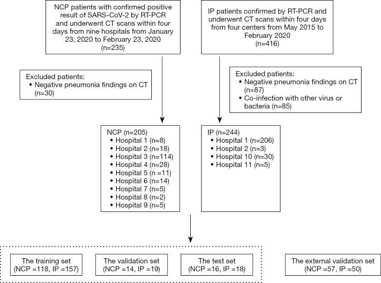
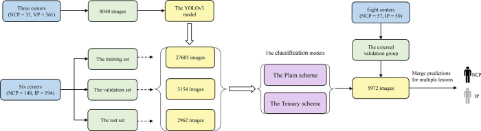
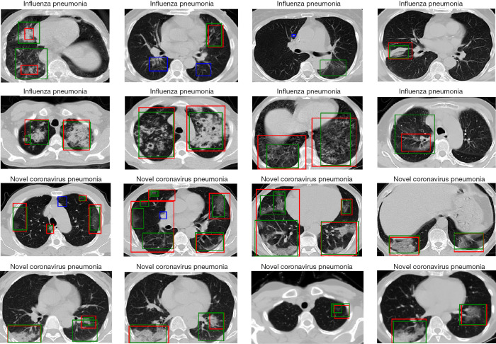
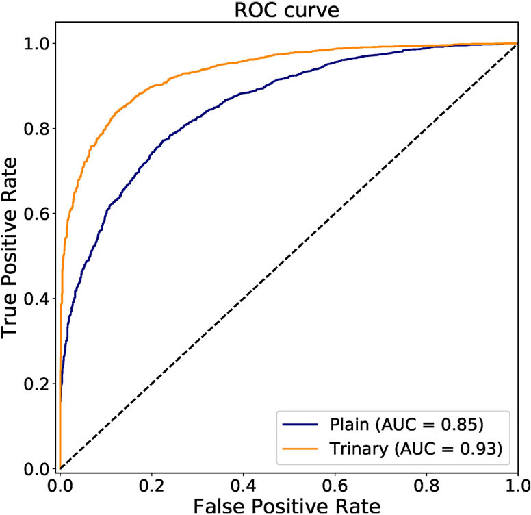
A total of 148 confirmed NCP patients [80 male; median age, 51.5 years; interquartile range (IQR), 42.5-63.0 years] treated in 4 NCP designated hospitals between January 11, 2020 and February 23, 2020 were retrospectively enrolled as a training cohort, along with 194 confirmed IP patients (112 males; median age, 65.0 years; IQR, 55.0-78.0 years) treated in 5 hospitals from May 2015 to February 2020. An external validation set comprising 57 NCP patients and 50 IP patients from 8 hospitals was also enrolled. Two deep learning schemes (the Trinary scheme and the Plain scheme) were developed and compared using receiver operating characteristic (ROC) curves.

RESULTS

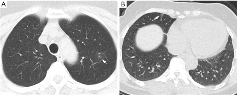
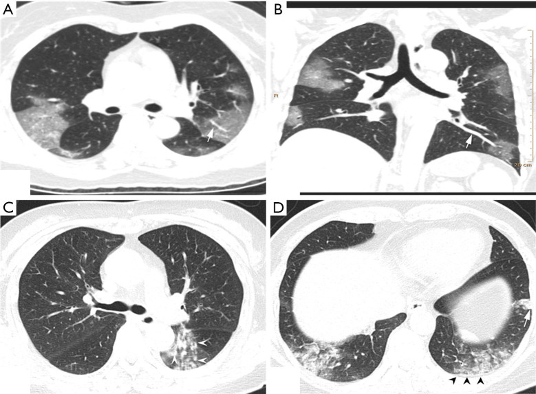
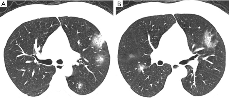
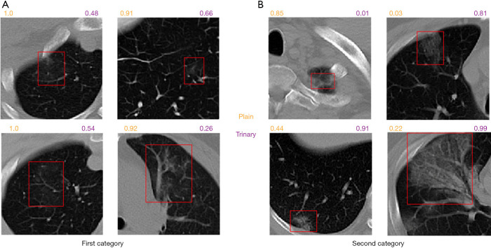
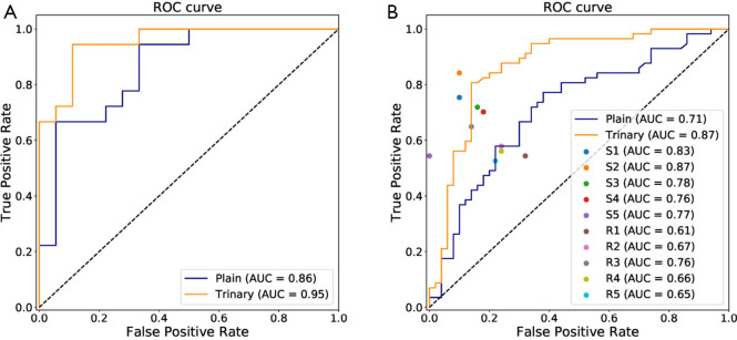
Of the NCP lesions, 96.6% were >1 cm and 76.8% were of a density <-500 Hu, indicating them to have less consolidation than IP lesions, which had nodules ranging from 5-10 mm. The Trinary scheme accurately distinguished NCP from IP lesions, with an area under the curve (AUC) of 0.93. For patient-level classification in the external validation set, the Trinary scheme outperformed the Plain scheme (AUC: 0.87 . 0.71) and achieved human specialist-level performance.

CONCLUSIONS

Our study has potentially provided an accurate tool on chest CT for early diagnosis of NCP with high transferability and showed high efficiency in differentiating between NCP and IP; these findings could help to reduce misdiagnosis and contain the pandemic transmission.

摘要

背景

胸部计算机断层扫描(CT)已被发现对新型冠状病毒肺炎(NCP)早期诊断具有高敏感性,在当前疫情期间,这使其相对于核酸检测具有优势。在本研究中,我们旨在开发并验证一个基于胸部CT图像的集成深度学习框架,用于自动检测NCP,尤其着重于将NCP与流感肺炎(IP)区分开来。

方法

回顾性纳入2020年1月11日至2020年2月23日期间在4家NCP定点医院接受治疗的148例确诊NCP患者[80例男性;中位年龄51.5岁;四分位间距(IQR),42.5 - 63.0岁]作为训练队列,以及2015年5月至2020年2月期间在5家医院接受治疗的194例确诊IP患者(112例男性;中位年龄65.0岁;IQR,55.0 - 78.0岁)。还纳入了一个由来自8家医院的57例NCP患者和50例IP患者组成的外部验证集。使用受试者操作特征(ROC)曲线开发并比较了两种深度学习方案(三元方案和普通方案)。

结果

NCP病灶中,96.6%大于1 cm,76.8%的密度小于 - 500 Hu,表明其实变程度低于IP病灶,IP病灶有5 - 10 mm的结节。三元方案能准确区分NCP与IP病灶,曲线下面积(AUC)为0.93。在外部验证集中进行患者水平分类时,三元方案优于普通方案(AUC:0.87对0.71),并达到了人类专家水平的性能。

结论

我们的研究可能为胸部CT早期诊断NCP提供了一种具有高可转移性的准确工具,并在区分NCP和IP方面显示出高效率;这些发现有助于减少误诊并遏制疫情传播。

https://cdn.ncbi.nlm.nih.gov/pmc/blobs/e245/7867927/7b9a8669cd1a/atm-09-02-111-f10.jpg
https://cdn.ncbi.nlm.nih.gov/pmc/blobs/e245/7867927/85d9a6e17783/atm-09-02-111-f1.jpg
https://cdn.ncbi.nlm.nih.gov/pmc/blobs/e245/7867927/041b04c42165/atm-09-02-111-f2.jpg
https://cdn.ncbi.nlm.nih.gov/pmc/blobs/e245/7867927/68a8b9eea250/atm-09-02-111-f3.jpg
https://cdn.ncbi.nlm.nih.gov/pmc/blobs/e245/7867927/d22a5cdba449/atm-09-02-111-f4.jpg
https://cdn.ncbi.nlm.nih.gov/pmc/blobs/e245/7867927/7edb6f93bb11/atm-09-02-111-f5.jpg
https://cdn.ncbi.nlm.nih.gov/pmc/blobs/e245/7867927/33bbdaef7994/atm-09-02-111-f6.jpg
https://cdn.ncbi.nlm.nih.gov/pmc/blobs/e245/7867927/92517b86b435/atm-09-02-111-f7.jpg
https://cdn.ncbi.nlm.nih.gov/pmc/blobs/e245/7867927/49e51a1cdf03/atm-09-02-111-f8.jpg
https://cdn.ncbi.nlm.nih.gov/pmc/blobs/e245/7867927/ecc7f246dec2/atm-09-02-111-f9.jpg
https://cdn.ncbi.nlm.nih.gov/pmc/blobs/e245/7867927/7b9a8669cd1a/atm-09-02-111-f10.jpg

相似文献

1
Deep learning for differentiating novel coronavirus pneumonia and influenza pneumonia.用于区分新型冠状病毒肺炎和流感肺炎的深度学习
Ann Transl Med. 2021 Jan;9(2):111. doi: 10.21037/atm-20-5328.
2
Radiomics-based model for accurately distinguishing between severe acute respiratory syndrome associated coronavirus 2 (SARS-CoV-2) and influenza A infected pneumonia.基于影像组学的模型用于准确区分严重急性呼吸综合征冠状病毒2(SARS-CoV-2)和甲型流感感染性肺炎。
MedComm (2020). 2020 Aug 13;1(2):240-248. doi: 10.1002/mco2.14. eCollection 2020 Sep.
3
Differentiation of pulmonary sclerosing pneumocytoma from solid malignant pulmonary nodules by radiomic analysis on multiphasic CT.多期 CT 放射组学分析对肺硬化性血管平滑肌脂肪瘤与实性恶性肺结节的鉴别诊断
J Appl Clin Med Phys. 2021 Feb;22(2):158-164. doi: 10.1002/acm2.13154. Epub 2020 Dec 28.
4
Differential Diagnosis of COVID-19 Pneumonia From Influenza A (H1N1) Pneumonia Using a Model Based on Clinicoradiologic Features.基于临床放射学特征的模型对新型冠状病毒肺炎与甲型H1N1流感肺炎的鉴别诊断
Front Med (Lausanne). 2021 Jun 15;8:651556. doi: 10.3389/fmed.2021.651556. eCollection 2021.
5
A human-computer collaboration for COVID-19 differentiation: combining a radiomics model with deep learning and human auditing.一种用于COVID-19鉴别诊断的人机协作:将放射组学模型与深度学习及人工审核相结合。
Ann Palliat Med. 2021 Jul;10(7):7329-7339. doi: 10.21037/apm-20-2625. Epub 2021 Jul 9.
6
CT-based radiomics combined with signs: a valuable tool to help radiologist discriminate COVID-19 and influenza pneumonia.基于 CT 的放射组学结合征象:有助于放射科医生鉴别 COVID-19 和流感肺炎的有价值工具。
BMC Med Imaging. 2021 Feb 17;21(1):31. doi: 10.1186/s12880-021-00564-w.
7
The characteristics and clinical value of chest CT images of novel coronavirus pneumonia.新型冠状病毒肺炎的胸部 CT 图像特征及临床价值。
Clin Radiol. 2020 May;75(5):335-340. doi: 10.1016/j.crad.2020.03.002. Epub 2020 Mar 19.
8
2019 Novel Coronavirus-Infected Pneumonia on CT: A Feasibility Study of Few-Shot Learning for Computerized Diagnosis of Emergency Diseases.2019新型冠状病毒感染肺炎的CT表现:急诊疾病计算机诊断的少样本学习可行性研究
IEEE Access. 2020 Oct 22;8:194158-194165. doi: 10.1109/ACCESS.2020.3033069. eCollection 2020.
9
Clinical and computed tomographic imaging features of novel coronavirus pneumonia caused by SARS-CoV-2.SARS-CoV-2 引起的新型冠状病毒肺炎的临床和计算机断层扫描影像学特征。
J Infect. 2020 Apr;80(4):394-400. doi: 10.1016/j.jinf.2020.02.017. Epub 2020 Feb 25.
10
From community-acquired pneumonia to COVID-19: a deep learning-based method for quantitative analysis of COVID-19 on thick-section CT scans.从社区获得性肺炎到 COVID-19:一种基于深度学习的 CT 厚层扫描 COVID-19 定量分析方法。
Eur Radiol. 2020 Dec;30(12):6828-6837. doi: 10.1007/s00330-020-07042-x. Epub 2020 Jul 18.

引用本文的文献

1
Improve the performance of CT-based pneumonia classification via source data reweighting.通过重新加权源数据来提高基于 CT 的肺炎分类性能。
Sci Rep. 2023 Jun 9;13(1):9401. doi: 10.1038/s41598-023-35938-3.
2
Artificial intelligence for differentiating COVID-19 from other viral pneumonias on CT: comparative analysis of different models based on quantitative and radiomic approaches.基于定量和影像组学方法的不同模型对 CT 鉴别 COVID-19 与其他病毒性肺炎的人工智能:比较分析。
Eur Radiol Exp. 2023 Jan 24;7(1):3. doi: 10.1186/s41747-022-00317-6.
3
Nurse, Give Me the News! Understanding Support for and Opposition to a COVID-19 Health Screening System.

本文引用的文献

1
Stop Explaining Black Box Machine Learning Models for High Stakes Decisions and Use Interpretable Models Instead.停止为高风险决策解释黑箱机器学习模型,转而使用可解释模型。
Nat Mach Intell. 2019 May;1(5):206-215. doi: 10.1038/s42256-019-0048-x. Epub 2019 May 13.
2
A comparison of deep learning performance against health-care professionals in detecting diseases from medical imaging: a systematic review and meta-analysis.深度学习在医学影像疾病检测方面的性能与医疗保健专业人员的比较:系统评价和荟萃分析。
Lancet Digit Health. 2019 Oct;1(6):e271-e297. doi: 10.1016/S2589-7500(19)30123-2. Epub 2019 Sep 25.
3
A deep learning approach to characterize 2019 coronavirus disease (COVID-19) pneumonia in chest CT images.
护士,给我消息!了解对 COVID-19 健康筛查系统的支持和反对。
Int J Environ Res Public Health. 2023 Jan 9;20(2):1164. doi: 10.3390/ijerph20021164.
4
Diagnostic Performance in Differentiating COVID-19 from Other Viral Pneumonias on CT Imaging: Multi-Reader Analysis Compared with an Artificial Intelligence-Based Model.CT 影像鉴别新型冠状病毒肺炎与其他病毒性肺炎的诊断效能:多读者分析与基于人工智能模型的比较。
Tomography. 2022 Nov 25;8(6):2815-2827. doi: 10.3390/tomography8060235.
5
SARS-CoV-2: Has artificial intelligence stood the test of time.SARS-CoV-2:人工智能是否经受住了时间的考验。
Chin Med J (Engl). 2022 Aug 5;135(15):1792-1802. doi: 10.1097/CM9.0000000000002058.
6
Comparison of temporal evolution of computed tomography imaging features in COVID-19 and influenza infections in a multicenter cohort study.一项多中心队列研究中COVID-19与流感感染的计算机断层扫描成像特征的时间演变比较。
Eur J Radiol Open. 2022;9:100431. doi: 10.1016/j.ejro.2022.100431. Epub 2022 Jun 24.
7
External Validation of Deep Learning Algorithms for Radiologic Diagnosis: A Systematic Review.用于放射诊断的深度学习算法的外部验证:一项系统评价。
Radiol Artif Intell. 2022 May 4;4(3):e210064. doi: 10.1148/ryai.210064. eCollection 2022 May.
8
ADA-COVID: Adversarial Deep Domain Adaptation-Based Diagnosis of COVID-19 from Lung CT Scans Using Triplet Embeddings.ADA-COVID:基于三重嵌入的对抗性深度域自适应的 COVID-19 肺部 CT 扫描诊断。
Comput Intell Neurosci. 2022 Feb 8;2022:2564022. doi: 10.1155/2022/2564022. eCollection 2022.
9
Clinical characteristics of patients with confirmed and suspected COVID-19.确诊和疑似新冠肺炎患者的临床特征
Am J Transl Res. 2021 Nov 15;13(11):12897-12904. eCollection 2021.
10
Artificial intelligence for imaging-based COVID-19 detection: Systematic review comparing added value of AI versus human readers.基于影像的 COVID-19 检测人工智能:比较 AI 与人工读者增值的系统评价。
Eur J Radiol. 2021 Dec;145:110028. doi: 10.1016/j.ejrad.2021.110028. Epub 2021 Nov 16.
深度学习在胸部 CT 图像中对 2019 冠状病毒病(COVID-19)肺炎的特征描述
Eur Radiol. 2020 Dec;30(12):6517-6527. doi: 10.1007/s00330-020-07044-9. Epub 2020 Jul 2.
4
A fully automatic deep learning system for COVID-19 diagnostic and prognostic analysis.一种用于 COVID-19 诊断和预后分析的全自动深度学习系统。
Eur Respir J. 2020 Aug 6;56(2). doi: 10.1183/13993003.00775-2020. Print 2020 Aug.
5
Severity and outcomes of influenza-related pneumonia in type A and B strains in China, 2013-2019.2013-2019 年中国 A 型和 B 型流感相关肺炎的严重程度和结局。
Infect Dis Poverty. 2020 Apr 22;9(1):42. doi: 10.1186/s40249-020-00655-w.
6
Laboratory data analysis of novel coronavirus (COVID-19) screening in 2510 patients.2510 例新型冠状病毒(COVID-19)筛查的实验室数据分析。
Clin Chim Acta. 2020 Aug;507:94-97. doi: 10.1016/j.cca.2020.04.018. Epub 2020 Apr 18.
7
High Contagiousness and Rapid Spread of Severe Acute Respiratory Syndrome Coronavirus 2.高传染性和严重急性呼吸综合征冠状病毒 2 的快速传播。
Emerg Infect Dis. 2020 Jul;26(7):1470-1477. doi: 10.3201/eid2607.200282. Epub 2020 Jun 21.
8
Coronavirus Disease 2019 and Influenza 2019-2020.2019年冠状病毒病与2019 - 2020年流感
JAMA. 2020 Mar 24;323(12):1122. doi: 10.1001/jama.2020.2633.
9
Using Artificial Intelligence to Detect COVID-19 and Community-acquired Pneumonia Based on Pulmonary CT: Evaluation of the Diagnostic Accuracy.基于肺部 CT 的人工智能检测 COVID-19 和社区获得性肺炎:诊断准确性评估。
Radiology. 2020 Aug;296(2):E65-E71. doi: 10.1148/radiol.2020200905. Epub 2020 Mar 19.
10
Detection of SARS-CoV-2 in Different Types of Clinical Specimens.SARS-CoV-2 在不同类型临床标本中的检测。
JAMA. 2020 May 12;323(18):1843-1844. doi: 10.1001/jama.2020.3786.