Curran R C, Jones E L
Clin Exp Immunol. 1977 Apr;28(1):103-15.
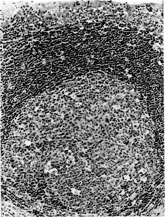
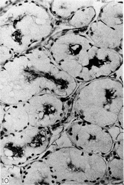
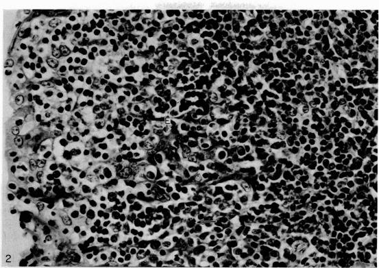
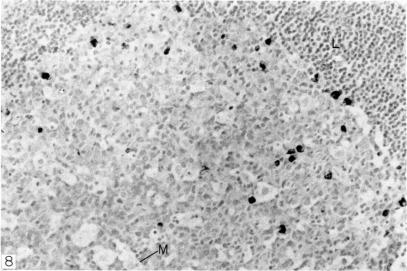
Intracellular immunoglobulin has been demonstrated in human palatine tonsils by the unlabelled antibody peroxidase-antiperoxidase complex (PAP) method in which rabbit antiserum to a range of human immunoglobulins (Igs) was linked to the PAP complex by an intermediate stage of swine antiserum to rabbit Ig. The effects of different methods of fixation and processing have been compared, formol-saline fixation giving the best results. The PAP technique proved greatly superior to the fluorescein isothiocyanate (FITC)-based technique, not only in sensitivity but in permitting study of the finer histological and cytological features. The lymphoid follicles are shown to have three distinct zones, two forming the follicle centre (zones (a) and (b)), and the third (zone (c)) the lymphocyte cap. Ig synthesis appeared to begin in the cells in zone (b). IgG, IgA, IgM, IgE and IgD were present in all tonsils, with IgG predominating, confirming that the tonsil resembles lymph nodes more closely than it does alimentary lymphoid tissue. Some follicles contained more than one type of Ig. The tonsil appears to have a well-developed T-dependent area, the lymphoid follicles forming a B-cell area. The structure of the tonsil would seem to facilitate contact between its lymphoid tissue and antigens in the crypts, and it is postulated that some T cells within the crypt epithelium, after contact with antigen, may leave the tonsil by the efferent lymphatics and enter the peripheral circulation by the thoracic duct, whilst other primed T cells interact with B cells in the follicle centres. Some B cells may then start to synthesize immunoglobulin, whilst others become memory cells in the lymphocyte 'cap' of the follicle.
运用未标记抗体过氧化物酶-抗过氧化物酶复合物(PAP)法在人腭扁桃体中证实了细胞内免疫球蛋白的存在。该方法中,一系列针对人免疫球蛋白(Ig)的兔抗血清通过猪抗兔Ig血清这一中间阶段与PAP复合物相连。比较了不同固定和处理方法的效果,结果显示甲醛生理盐水固定效果最佳。PAP技术被证明在灵敏度上以及在对更精细的组织学和细胞学特征的研究方面都大大优于基于异硫氰酸荧光素(FITC)的技术。淋巴滤泡显示有三个不同区域,两个构成滤泡中心(区域(a)和(b)),第三个(区域(c))是淋巴细胞帽。Ig合成似乎始于区域(b)的细胞。所有扁桃体中均存在IgG、IgA、IgM、IgE和IgD,其中IgG占主导,这证实扁桃体与淋巴结的相似性高于与消化道淋巴组织的相似性。一些滤泡含有不止一种类型的Ig。扁桃体似乎有一个发育良好的T细胞依赖区,淋巴滤泡构成B细胞区。扁桃体的结构似乎便于其淋巴组织与隐窝中的抗原接触,据推测,隐窝上皮内的一些T细胞在与抗原接触后,可能通过输出淋巴管离开扁桃体,并经胸导管进入外周循环,而其他致敏T细胞则在滤泡中心与B细胞相互作用。然后一些B细胞可能开始合成免疫球蛋白,而其他B细胞则在滤泡的淋巴细胞“帽”中成为记忆细胞。