Ravi Shravanthy, Santhanakrishnan Muthukumar
Department of Peridontology, Faculty of Dental Sciences, Sri Ramachandra Institute of Higher Education and Research, Porur, Chennai, 600116 India.
Biomater Res. 2020 Sep 11;24:16. doi: 10.1186/s40824-020-00193-4. eCollection 2020.
Platelet concentrates have been popularly used in regenerative periodontal therapy as they are autologous in origin and they provide a supernatural concentration of platelets, growth factors and leukocytes. The release profile of various growth factors is considered important during the various phases of wound healing with the most important being the inflammatory phase where the release of the growth factors help in recruitment of cells and in collagen production. With the more recent modifications of PRF namely A-PRF and T-PRF, the mechanical and chemical degradation properties have also improved. The aim of the present study was to correlate the release profile of PDGF-AA from various forms of platelet concentrates (L-PRF, A-PRF, T-PRF) based on their mechanical and chemical properties.
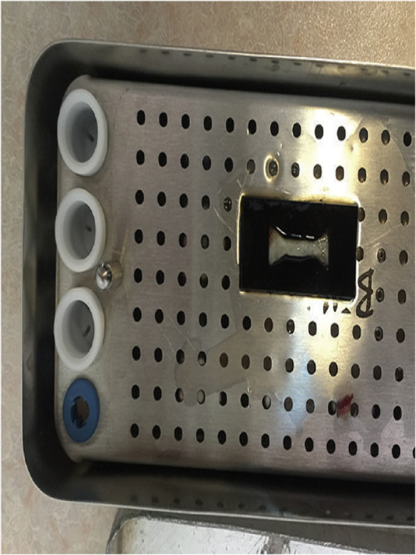
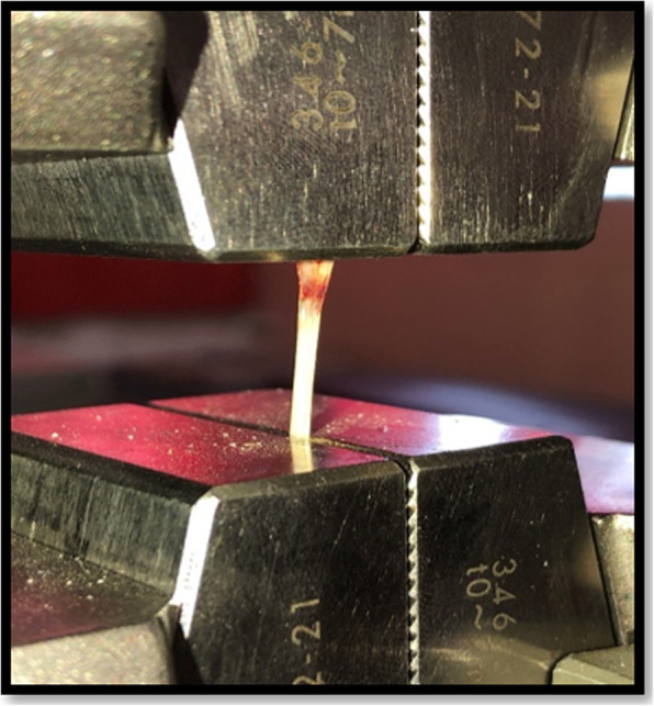
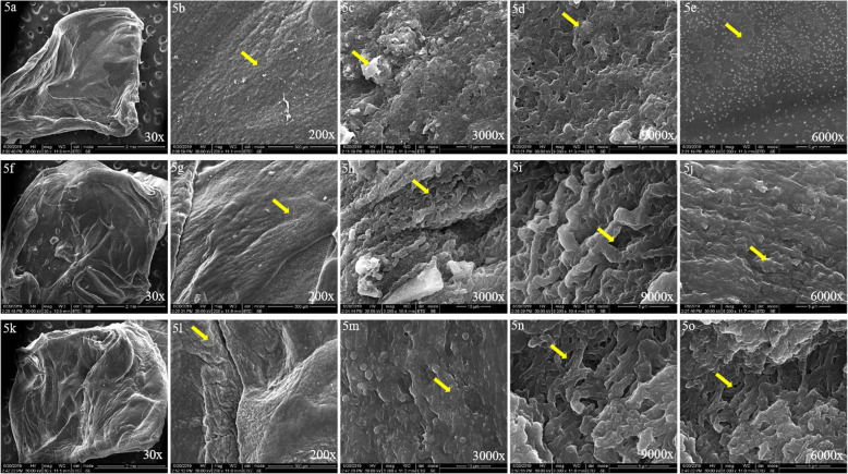
Blood samples were drawn from 2 male and 3 female systemically healthy patients between 20 and 25 years of age who were about to undergo periodontal regeneration for PRF preparation. The blood sample was immediately centrifuged using a table top centrifuge (Remi R4C) at 1060 rpm (208 x g) for 14 min for A-PRF preparation, 1960 rpm (708 x g) for 12 min for L-PRF preparation and 1960 rpm (708 x g) for 12 min in titanium tubes for T-PRF preparation. Tensile test was performed using universal testing machine. The in vitro degradation test of the prepared PRF membranes were conducted by placing the PRF membrane in 10 ml of pH 7.4 PBS on an orbital shaker set at 50 rpm. SEM evaluation of the PRF membrane was done under both low and high magnification. In order to determine the amount of released growth factor PDGF-AA at 15 min, 60 min, 8 h, 1 day, 3 days, and 10 days, samples were placed into a shaking incubator at 37 °C to allow for growth factor release into the culture media.
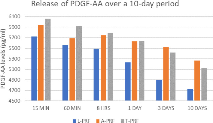
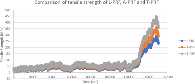
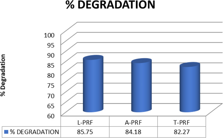
On comparing the three PRF membranes, it was found that T-PRF contained the maximum tensile strength (404.61 ± 5.92 MPa) and modulus of elasticity (151.9 ± 6.92 MPa). Statistically significant differences between the three groups were found on comparing the groups for their mechanical properties. In the degradation test, it was found that the maximum amount of degradation was found in L-PRF (85.75%), followed by A-PRF (84.18%) and the least was found in T-PRF (82.27%). T-PRF released the highest amount of PDGF-AA (6060.4 pg/ml) at early time points when compared to A-PRF (5935.3 pg/ml). While T-PRF had rapid release of PDGF-AA, A-PRF had a sustained release of growth factors released at later time points.
Results from the present study indicate that A-PRF is the most favourable form of platelet concentrate in regenerative periodontal therapy as it has a sustained release of growth factors over time.
血小板浓缩物因其来源为自体且能提供超自然浓度的血小板、生长因子和白细胞,已广泛应用于牙周再生治疗。在伤口愈合的各个阶段,各种生长因子的释放模式被认为很重要,其中最重要的是炎症阶段,生长因子的释放在细胞募集和胶原蛋白生成中起作用。随着PRF(富血小板纤维蛋白)的最新改进,即A-PRF和T-PRF,其机械和化学降解特性也有所改善。本研究的目的是根据不同形式血小板浓缩物(L-PRF、A-PRF、T-PRF)的机械和化学性质,关联其PDGF-AA(血小板衍生生长因子AA)的释放模式。
从20至25岁、全身健康的2名男性和3名女性患者中采集血样,这些患者即将接受牙周再生治疗以制备PRF。血样立即使用台式离心机(Remi R4C)以1060转/分钟(208×g)离心14分钟制备A-PRF,以1960转/分钟(708×g)离心12分钟制备L-PRF,在钛管中以1960转/分钟(708×g)离心12分钟制备T-PRF。使用万能试验机进行拉伸试验。将制备的PRF膜置于10毫升pH值为7.4的PBS中,在设置为50转/分钟的轨道振荡器上进行体外降解试验。在低倍和高倍显微镜下对PRF膜进行扫描电子显微镜(SEM)评估。为了测定在15分钟、60分钟、8小时、1天、3天和10天时释放的生长因子PDGF-AA的量,将样品置于37°C的振荡培养箱中,以使生长因子释放到培养基中。
比较三种PRF膜发现,T-PRF的拉伸强度最高(404.61±5.92兆帕),弹性模量最大(151.9±6.92兆帕)。比较三组的机械性能时发现三组之间存在统计学显著差异。在降解试验中,发现L-PRF的降解量最大(85.75%),其次是A-PRF(84.18%),T-PRF的降解量最少(82.27%)。与A-PRF(5935.3皮克/毫升)相比,T-PRF在早期时间点释放的PDGF-AA量最高(6060.4皮克/毫升)。虽然T-PRF的PDGF-AA释放迅速,但A-PRF在后期时间点释放的生长因子具有持续释放的特点。
本研究结果表明,A-PRF是牙周再生治疗中最有利的血小板浓缩物形式,因为它的生长因子随时间持续释放。