Institute of Diagnostic and Interventional Radiology and Neuroradiology, University Hospital Essen, Essen, Germany.
Department of General, Visceral and Transplantation Surgery, University Hospital Essen, Essen, Germany.
Eur Radiol. 2021 Apr;31(4):1795-1804. doi: 10.1007/s00330-020-07147-3. Epub 2020 Sep 18.
Body tissue composition is a long-known biomarker with high diagnostic and prognostic value not only in cardiovascular, oncological, and orthopedic diseases but also in rehabilitation medicine or drug dosage. In this study, the aim was to develop a fully automated, reproducible, and quantitative 3D volumetry of body tissue composition from standard CT examinations of the abdomen in order to be able to offer such valuable biomarkers as part of routine clinical imaging.
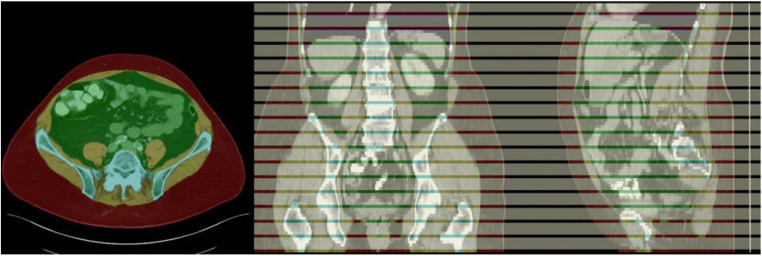
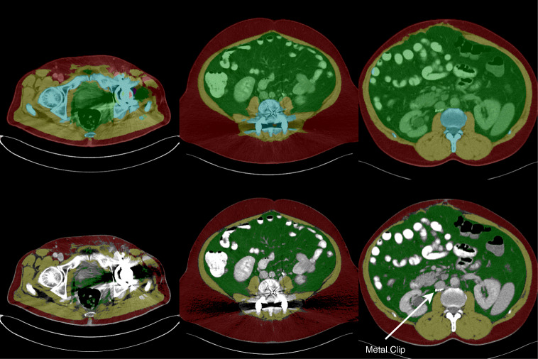
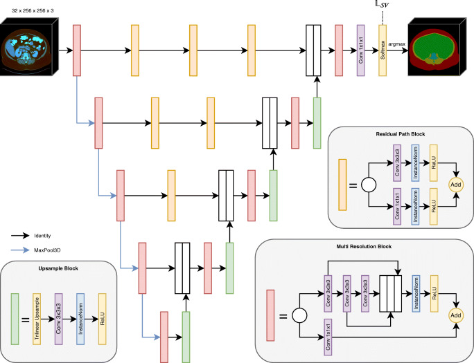
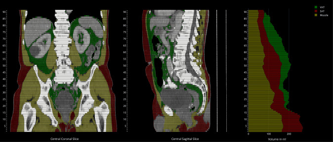
Therefore, an in-house dataset of 40 CTs for training and 10 CTs for testing were fully annotated on every fifth axial slice with five different semantic body regions: abdominal cavity, bones, muscle, subcutaneous tissue, and thoracic cavity. Multi-resolution U-Net 3D neural networks were employed for segmenting these body regions, followed by subclassifying adipose tissue and muscle using known Hounsfield unit limits.
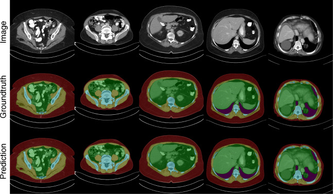
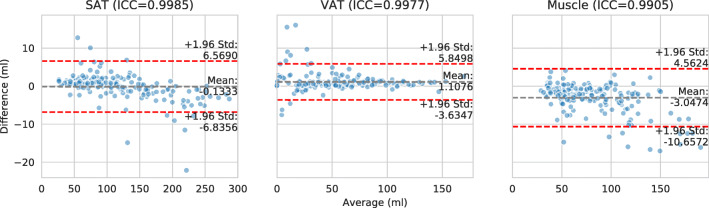
The Sørensen Dice scores averaged over all semantic regions was 0.9553 and the intra-class correlation coefficients for subclassified tissues were above 0.99.
Our results show that fully automated body composition analysis on routine CT imaging can provide stable biomarkers across the whole abdomen and not just on L3 slices, which is historically the reference location for analyzing body composition in the clinical routine.
• Our study enables fully automated body composition analysis on routine abdomen CT scans. • The best segmentation models for semantic body region segmentation achieved an averaged Sørensen Dice score of 0.9553. • Subclassified tissue volumes achieved intra-class correlation coefficients over 0.99.
人体组织成分是一种由来已久的生物标志物,不仅在心血管、肿瘤学和骨科疾病中具有很高的诊断和预后价值,而且在康复医学或药物剂量中也是如此。本研究旨在开发一种全自动、可重复、定量的腹部标准 CT 检查的人体组织成分 3D 容积测量方法,以便能够提供这些有价值的生物标志物作为常规临床成像的一部分。
因此,我们使用了一个内部的 40 个 CT 训练数据集和 10 个 CT 测试数据集,对每 5 个轴向切片进行了完全注释,共涉及 5 个不同的语义体区:腹腔、骨骼、肌肉、皮下组织和胸腔。使用多分辨率 U-Net 3D 神经网络对这些体区进行分割,然后使用已知的 Hounsfield 单位界限对脂肪组织和肌肉进行亚分类。
所有语义区域的平均 Sørensen Dice 评分均为 0.9553,亚分类组织的组内相关系数均高于 0.99。
我们的结果表明,在常规 CT 成像上进行全自动的人体成分分析可以提供整个腹部的稳定生物标志物,而不仅仅是在 L3 切片上,这在临床常规中一直是分析人体成分的参考位置。
我们的研究能够在常规腹部 CT 扫描上进行全自动的人体成分分析。
用于语义体区分割的最佳分割模型的平均 Sørensen Dice 评分达到 0.9553。
亚分类组织体积的组内相关系数超过 0.99。