From the Department of Radiology, Nationwide Children's Hospital, 700 Children's Dr, Columbus, OH 43235 (H.H.H., M.A.S.); Department of Radiology, University of Texas Southwestern Medical Center, Dallas, Tex (T.Y.); Department of Radiology (M.R.B., J.S.), Department of Medicine, Division of Gastroenterology (M.R.B.), and Center for Advanced Magnetic Resonance Development (M.R.B., J.S.), Duke University Medical Center, Durham, NC; Liver Imaging Group, Department of Radiology, University of California San Diego, San Diego, Calif (C.B.S., M.S.M., W.C.H., G.H.); Departments of Radiology (D.H., J.H.B., S.B.R.), Medical Physics (D.H., E.F.J., S.B.R.), Biomedical Engineering (S.B.R.), Medicine (S.B.R.), and Emergency Medicine (S.B.R.), University of Wisconsin, Madison, Wis; Department of Radiology, University of Michigan, Ann Arbor, Mich (D.M., T.L.C.); Department of Radiology, Children's Hospital of Philadelphia, Philadelphia, Pa (S.D.S.); Department of Radiology, Mayo Clinic, Rochester, Minn (Y.S.); Department of Radiology, Cincinnati Children's Hospital Medical Center, Cincinnati, Ohio (J.A.T., A.T.T.); Department of Radiology, University of Cincinnati College of Medicine, Cincinnati, Ohio (J.A.T., A.T.T.); Department of Quantitative Health Science, Cleveland Clinic Foundation, Cleveland, Ohio (N.O.); and Calimetrix, LLC, Madison, Wis (J.H.B.).
Radiology. 2021 Mar;298(3):640-651. doi: 10.1148/radiol.2021202912. Epub 2021 Jan 19.
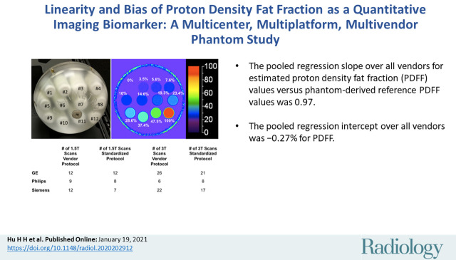
Background Proton density fat fraction (PDFF) estimated by using chemical shift-encoded (CSE) MRI is an accepted imaging biomarker of hepatic steatosis. This work aims to promote standardized use of CSE MRI to estimate PDFF. Purpose To assess the accuracy of CSE MRI methods for estimating PDFF by determining the linearity and range of bias observed in a phantom. Materials and Methods In this prospective study, a commercial phantom with 12 vials of known PDFF values were shipped across nine U.S. centers. The phantom underwent 160 independent MRI examinations on 27 1.5-T and 3.0-T systems from three vendors. Two three-dimensional CSE MRI protocols with minimal T1 bias were included: vendor and standardized. Each vendor's confounder-corrected complex or hybrid magnitude-complex based reconstruction algorithm was used to generate PDFF maps in both protocols. The Siemens reconstruction required a configuration change to correct for water-fat swaps in the phantom. The MRI PDFF values were compared with the known PDFF values by using linear regression with mixed-effects modeling. The 95% CIs were calculated for the regression slope (ie, proportional bias) and intercept (ie, constant bias) and compared with the null hypothesis (slope = 1, intercept = 0). Results Pooled regression slope for estimated PDFF values versus phantom-derived reference PDFF values was 0.97 (95% CI: 0.96, 0.98) in the biologically relevant 0%-47.5% PDFF range. The corresponding pooled intercept was -0.27% (95% CI: -0.50%, -0.05%). Across vendors, slope ranges were 0.86-1.02 (vendor protocols) and 0.97-1.0 (standardized protocol) at 1.5 T and 0.91-1.01 (vendor protocols) and 0.87-1.01 (standardized protocol) at 3.0 T. The intercept ranges (absolute PDFF percentage) were -0.65% to 0.18% (vendor protocols) and -0.69% to -0.17% (standardized protocol) at 1.5 T and -0.48% to 0.10% (vendor protocols) and -0.78% to -0.21% (standardized protocol) at 3.0 T. Conclusion Proton density fat fraction estimation derived from three-dimensional chemical shift-encoded MRI in a commercial phantom was accurate across vendors, imaging centers, and field strengths, with use of the vendors' product acquisition and reconstruction software. © RSNA, 2021 See also the editorial by Dyke in this issue.
背景 利用化学位移编码(CSE)MRI 估计的质子密度脂肪分数(PDFF)是肝脂肪变性的公认影像学生物标志物。本研究旨在通过确定体模中观察到的线性和偏倚范围,促进 CSE MRI 估计 PDFF 的标准化使用。目的 评估 CSE MRI 方法估计 PDFF 的准确性,方法是确定在商业体模中测量得到的与已知 PDFF 值的线性关系和偏倚范围。材料与方法 本前瞻性研究中,一个装有 12 个已知 PDFF 值小瓶的商用体模被运往美国 9 个中心。体模在 3 家供应商的 27 台 1.5-T 和 3.0-T 系统上进行了 160 次独立的 MRI 检查。包括两种具有最小 T1 偏倚的三维 CSE MRI 方案:供应商和标准化。每个供应商的混杂因素校正复杂或混合幅度复杂的基于重建算法用于在两个方案中生成 PDFF 图。西门子重建需要进行配置更改,以纠正体模中的水 - 脂肪交换。通过混合效应模型的线性回归,将 MRI 的 PDFF 值与已知 PDFF 值进行比较。计算回归斜率(即比例偏差)和截距(即恒定偏差)的 95%置信区间,并与零假设(斜率=1,截距=0)进行比较。结果 在 0%-47.5%的生物相关 PDFF 范围内,估计的 PDFF 值与体模衍生参考 PDFF 值的汇总回归斜率为 0.97(95%CI:0.96,0.98)。相应的汇总截距为-0.27%(95%CI:-0.50%,-0.05%)。在不同供应商中,1.5-T 时斜率范围为 0.86-1.02(供应商方案)和 0.97-1.0(标准化方案),3.0-T 时斜率范围为 0.91-1.01(供应商方案)和 0.87-1.01(标准化方案)。1.5-T 时截距范围(绝对 PDFF%)为-0.65%至 0.18%(供应商方案)和-0.69%至-0.17%(标准化方案),3.0-T 时截距范围为-0.48%至 0.10%(供应商方案)和-0.78%至-0.21%(标准化方案)。结论 在商业体模中,使用三维化学位移编码 MRI 从三个供应商的产品采集和重建软件中准确地估计了质子密度脂肪分数,结果在不同供应商、成像中心和场强中具有可重复性。 ©RSNA,2021 另见本期 Dyke 的社论。