• 文献检索
  • 文档翻译
  • 深度研究
  • 学术资讯
  • Suppr Zotero 插件Zotero 插件
  • 邀请有礼
  • 套餐&价格
  • 历史记录
应用&插件
Suppr Zotero 插件Zotero 插件浏览器插件Mac 客户端Windows 客户端微信小程序
定价
高级版会员购买积分包购买API积分包
服务
文献检索文档翻译深度研究API 文档MCP 服务
关于我们
关于 Suppr公司介绍联系我们用户协议隐私条款
关注我们

Suppr 超能文献

核心技术专利:CN118964589B侵权必究
粤ICP备2023148730 号-1Suppr @ 2026

文献检索

告别复杂PubMed语法,用中文像聊天一样搜索,搜遍4000万医学文献。AI智能推荐,让科研检索更轻松。

立即免费搜索

文件翻译

保留排版,准确专业,支持PDF/Word/PPT等文件格式,支持 12+语言互译。

免费翻译文档

深度研究

AI帮你快速写综述,25分钟生成高质量综述,智能提取关键信息,辅助科研写作。

立即免费体验

经碱水解改性的3D打印聚己内酯/羟基磷灰石支架增强体外成骨作用

3D-Printed Poly(ε-Caprolactone)/Hydroxyapatite Scaffolds Modified with Alkaline Hydrolysis Enhance Osteogenesis In Vitro.

作者信息

Park Sangbae, Kim Jae Eun, Han Jinsub, Jeong Seung, Lim Jae Woon, Lee Myung Chul, Son Hyunmok, Kim Hong Bae, Choung Yun-Hoon, Seonwoo Hoon, Chung Jong Hoon, Jang Kyoung-Je

机构信息

Department of Biosystems & Biomaterials Science and Engineering, Seoul National University, Seoul 08826, Korea.

Department of Biosystems Engineering, Seoul National University, Seoul 08826, Korea.

出版信息

Polymers (Basel). 2021 Jan 14;13(2):257. doi: 10.3390/polym13020257.

DOI:10.3390/polym13020257
PMID:33466736
原文链接:https://pmc.ncbi.nlm.nih.gov/articles/PMC7830212/
Abstract

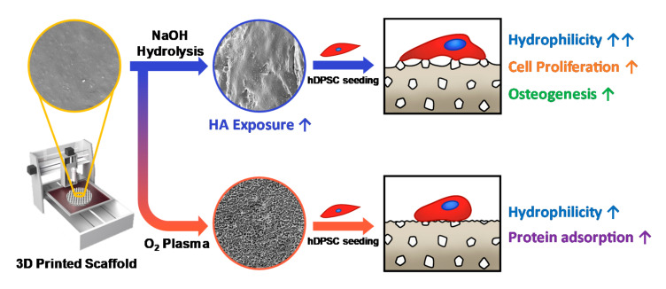
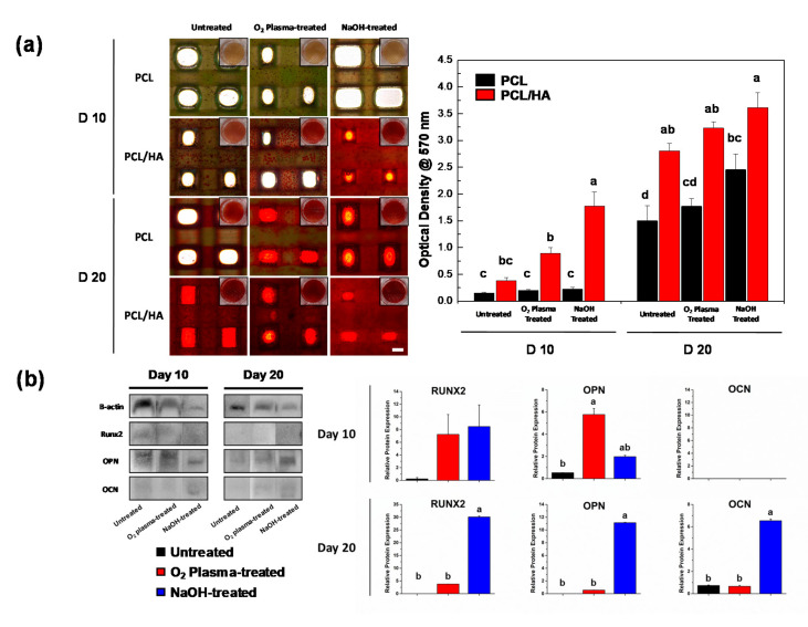
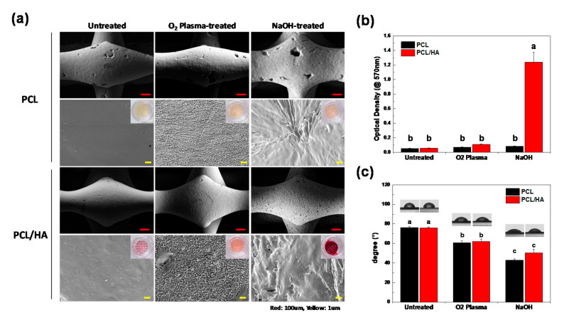
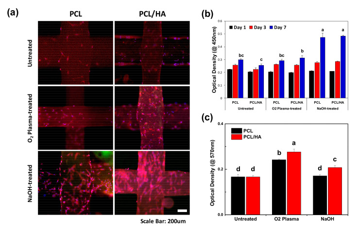
The 3D-printed bioactive ceramic incorporated Poly(ε-caprolactone) (PCL) scaffolds show great promise as synthetic bone graft substitutes. However, 3D-printed scaffolds still lack adequate surface properties for cells to be attached to them. In this study, we modified the surface characteristics of 3D-printed poly(ε-caprolactone)/hydroxyapatite scaffolds using O2 plasma and sodium hydroxide. The surface property of the alkaline hydrolyzed and O2 plasma-treated PCL/HA scaffolds were evaluated using field-emission scanning microscopy (FE-SEM), Alizarin Red S (ARS) staining, and water contact angle analysis, respectively. The in vitro behavior of the scaffolds was investigated using human dental pulp-derived stem cells (hDPSCs). Cell proliferation of hDPSCs on the scaffolds was evaluated via immunocytochemistry (ICC) and water-soluble tetrazolium salt (WST-1) assay. Osteogenic differentiation of hDPSCs on the scaffolds was further investigated using ARS staining and Western blot analysis. The result of this study shows that alkaline treatment is beneficial for exposing hydroxyapatite particles embedded in the scaffolds compared to O2 plasma treatment, which promotes cell proliferation and differentiation of hDPSCs.

摘要

3D打印的生物活性陶瓷增强聚己内酯(PCL)支架作为合成骨移植替代物显示出巨大的潜力。然而,3D打印的支架仍然缺乏足够的表面特性以供细胞附着。在本研究中,我们使用氧气等离子体和氢氧化钠对3D打印的聚己内酯/羟基磷灰石支架的表面特性进行了改性。分别使用场发射扫描显微镜(FE-SEM)、茜素红S(ARS)染色和水接触角分析对经碱水解和氧气等离子体处理的PCL/HA支架的表面性能进行了评估。使用人牙髓来源的干细胞(hDPSCs)研究了支架的体外行为。通过免疫细胞化学(ICC)和水溶性四唑盐(WST-1)测定评估了hDPSCs在支架上的细胞增殖。使用ARS染色和蛋白质印迹分析进一步研究了hDPSCs在支架上的成骨分化。本研究结果表明,与氧气等离子体处理相比,碱处理有利于暴露嵌入支架中的羟基磷灰石颗粒,从而促进hDPSCs的细胞增殖和分化。

https://cdn.ncbi.nlm.nih.gov/pmc/blobs/2c7d/7830212/2787d4d48804/polymers-13-00257-g004.jpg
https://cdn.ncbi.nlm.nih.gov/pmc/blobs/2c7d/7830212/e1c7a1e6c284/polymers-13-00257-g001.jpg
https://cdn.ncbi.nlm.nih.gov/pmc/blobs/2c7d/7830212/c382ba9f487c/polymers-13-00257-g002.jpg
https://cdn.ncbi.nlm.nih.gov/pmc/blobs/2c7d/7830212/ccca744d6c95/polymers-13-00257-g003.jpg
https://cdn.ncbi.nlm.nih.gov/pmc/blobs/2c7d/7830212/2787d4d48804/polymers-13-00257-g004.jpg

相似文献

1
3D-Printed Poly(ε-Caprolactone)/Hydroxyapatite Scaffolds Modified with Alkaline Hydrolysis Enhance Osteogenesis In Vitro.经碱水解改性的3D打印聚己内酯/羟基磷灰石支架增强体外成骨作用
Polymers (Basel). 2021 Jan 14;13(2):257. doi: 10.3390/polym13020257.
2
Enhanced osteogenic activity by MC3T3-E1 pre-osteoblasts on chemically surface-modified poly(ε-caprolactone) 3D-printed scaffolds compared to RGD immobilized scaffolds.MC3T3-E1 前成骨细胞在化学表面改性聚己内酯 3D 打印支架上的增强成骨活性优于 RGD 固定支架。
Biomed Mater. 2018 Nov 13;14(1):015008. doi: 10.1088/1748-605X/aaeb82.
3
Enhanced Osteogenic Differentiation of Periodontal Ligament Stem Cells Using a Graphene Oxide-Coated Poly(ε-caprolactone) Scaffold.使用氧化石墨烯包覆的聚己内酯支架增强牙周膜干细胞的成骨分化
Polymers (Basel). 2021 Mar 5;13(5):797. doi: 10.3390/polym13050797.
4
A Comparison of the Effects of Silica and Hydroxyapatite Nanoparticles on Poly(ε-caprolactone)-Poly(ethylene glycol)-Poly(ε-caprolactone)/Chitosan Nanofibrous Scaffolds for Bone Tissue Engineering.二氧化硅和羟基磷灰石纳米颗粒对用于骨组织工程的聚(ε-己内酯)-聚(乙二醇)-聚(ε-己内酯)/壳聚糖纳米纤维支架的影响比较
Tissue Eng Regen Med. 2018 Aug 14;15(6):735-750. doi: 10.1007/s13770-018-0140-z. eCollection 2018 Dec.
5
Enhanced osteogenic differentiation of stem cells by 3D printed PCL scaffolds coated with collagen and hydroxyapatite.3D 打印的聚己内酯支架表面涂胶原和羟基磷灰石增强干细胞的成骨分化。
Sci Rep. 2022 Jul 20;12(1):12359. doi: 10.1038/s41598-022-15602-y.
6
Effects of 3-dimensional Bioprinting Alginate/Gelatin Hydrogel Scaffold Extract on Proliferation and Differentiation of Human Dental Pulp Stem Cells.3D 生物打印海藻酸钙/明胶水凝胶支架浸提液对人牙髓干细胞增殖和分化的影响。
J Endod. 2019 Jun;45(6):706-715. doi: 10.1016/j.joen.2019.03.004. Epub 2019 May 2.
7
Osteogenic potential of human dental pulp stem cells cultured onto poly-ε-caprolactone/poly (rotaxane) scaffolds.人牙髓干细胞在聚己内酯/聚(轮烷)支架上培养的成骨潜能。
Dent Mater. 2019 Dec;35(12):1740-1749. doi: 10.1016/j.dental.2019.08.109. Epub 2019 Sep 20.
8
Osteoinduction and proliferation of bone-marrow stromal cells in three-dimensional poly (ε-caprolactone)/ hydroxyapatite/collagen scaffolds.三维聚(ε-己内酯)/羟基磷灰石/胶原蛋白支架中骨髓基质细胞的骨诱导与增殖
J Transl Med. 2015 May 8;13:152. doi: 10.1186/s12967-015-0499-8.
9
Adhesion, proliferation and osteogenic differentiation of mesenchymal stem cells in 3D printed poly-ε-caprolactone/hydroxyapatite scaffolds combined with bone marrow clots.3D 打印聚己内酯/羟基磷灰石支架联合骨髓凝块对间充质干细胞的黏附、增殖和成骨分化。
Mol Med Rep. 2017 Oct;16(4):5078-5084. doi: 10.3892/mmr.2017.7266. Epub 2017 Aug 17.
10
Osteoregenerative Potential of 3D-Printed Poly -Caprolactone Tissue Scaffolds In Vitro Using Minimally Manipulative Expansion of Primary Human Bone Marrow Stem Cells.体外使用最小化操作的原代人骨髓基质干细胞扩增三维打印聚己内酯组织支架的成骨再生潜力。
Int J Mol Sci. 2023 Mar 3;24(5):4940. doi: 10.3390/ijms24054940.

引用本文的文献

1
Integrating Physical and Biochemical Cues for Muscle Engineering: Scaffolds and Graft Durability.整合物理和生化线索用于肌肉组织工程:支架与移植物的耐久性
Bioengineering (Basel). 2024 Dec 9;11(12):1245. doi: 10.3390/bioengineering11121245.
2
A Systematic Review of the Contribution of Additive Manufacturing toward Orthopedic Applications.增材制造对骨科应用贡献的系统评价
ACS Omega. 2024 Oct 25;9(44):44042-44075. doi: 10.1021/acsomega.4c04870. eCollection 2024 Nov 5.
3
Microfluidic Fabrication of Gelatin-Nano Hydroxyapatite Scaffolds for Enhanced Control of Pore Size Distribution and Osteogenic Differentiation of Dental Pulp Stem Cells.

本文引用的文献

1
Three-Dimensional High-Porosity Chitosan/Honeycomb Porous Carbon/Hydroxyapatite Scaffold with Enhanced Osteoinductivity for Bone Regeneration.具有增强成骨活性的三维高孔隙率壳聚糖/蜂窝状多孔碳/羟基磷灰石支架用于骨再生。
ACS Biomater Sci Eng. 2020 Jan 13;6(1):575-586. doi: 10.1021/acsbiomaterials.9b01381. Epub 2019 Dec 13.
2
3D printing of silk microparticle reinforced polycaprolactone scaffolds for tissue engineering applications.用于组织工程应用的丝素微粒增强聚己内酯支架的3D打印
Mater Sci Eng C Mater Biol Appl. 2021 Jan;118:111433. doi: 10.1016/j.msec.2020.111433. Epub 2020 Aug 25.
3
3D-Printing of Hierarchically Designed and Osteoconductive Bone Tissue Engineering Scaffolds.
用于增强牙髓干细胞孔径分布控制和成骨分化的明胶-纳米羟基磷灰石支架的微流体制备
Macromol Biosci. 2024 Dec;24(12):e2400279. doi: 10.1002/mabi.202400279. Epub 2024 Oct 10.
4
Polycaprolactone in Bone Tissue Engineering: A Comprehensive Review of Innovations in Scaffold Fabrication and Surface Modifications.聚己内酯在骨组织工程中的应用:支架制造与表面修饰创新的综合评述
J Funct Biomater. 2024 Aug 24;15(9):243. doi: 10.3390/jfb15090243.
5
3D printed scaffolds of biosilica and spongin from marine sponges: analysis of genotoxicity and cytotoxicity for bone tissue repair.来自海洋海绵的生物二氧化硅和海绵硬蛋白的3D打印支架:用于骨组织修复的遗传毒性和细胞毒性分析
Bioprocess Biosyst Eng. 2024 Sep;47(9):1483-1498. doi: 10.1007/s00449-024-03042-z. Epub 2024 Jun 13.
6
Advances in 3D bioprinting for regenerative medicine applications.用于再生医学应用的3D生物打印技术进展。
Regen Biomater. 2024 Mar 26;11:rbae033. doi: 10.1093/rb/rbae033. eCollection 2024.
7
Evaluation of Pore Size Graded Bone Scaffolds with Different Material Composition.不同材料组成的孔径分级骨支架的评估
3D Print Addit Manuf. 2024 Apr 1;11(2):e718-e730. doi: 10.1089/3dp.2022.0138. Epub 2024 Apr 16.
8
Mechanical properties, and biocompatibility assessment of biomimetic dual layered keratin/ hydroxyapatite scaffolds.仿生双层角蛋白/羟基磷灰石支架的力学性能及生物相容性评估
Front Bioeng Biotechnol. 2023 Dec 15;11:1304147. doi: 10.3389/fbioe.2023.1304147. eCollection 2023.
9
Effect of Pore Characteristics and Alkali Treatment on the Physicochemical and Biological Properties of a 3D-Printed Polycaprolactone Bone Scaffold.孔隙特征和碱处理对3D打印聚己内酯骨支架理化及生物学性能的影响
ACS Omega. 2023 Feb 16;8(8):7378-7394. doi: 10.1021/acsomega.2c05571. eCollection 2023 Feb 28.
10
Functional engineering strategies of 3D printed implants for hard tissue replacement.用于硬组织替代的3D打印植入物的功能工程策略。
Regen Biomater. 2022 Nov 24;10:rbac094. doi: 10.1093/rb/rbac094. eCollection 2023.
分层设计且具有骨传导性的骨组织工程支架的3D打印
Materials (Basel). 2020 Apr 13;13(8):1836. doi: 10.3390/ma13081836.
4
Preparation and characterization of PLA/PCL/HA composite scaffolds using indirect 3D printing for bone tissue engineering.采用间接 3D 打印技术制备 PLA/PCL/HA 复合支架用于骨组织工程。
Mater Sci Eng C Mater Biol Appl. 2019 Nov;104:109960. doi: 10.1016/j.msec.2019.109960. Epub 2019 Jul 6.
5
Modelling and optimization of NaOH-etched 3-D printed PCL for enhanced cellular attachment and growth with minimal loss of mechanical strength.NaOH 刻蚀 3D 打印 PCL 的建模与优化,以增强细胞黏附与生长,同时最小化机械强度损失。
Mater Sci Eng C Mater Biol Appl. 2019 May;98:602-611. doi: 10.1016/j.msec.2018.12.084. Epub 2019 Jan 4.
6
A strong, tough, and osteoconductive hydroxyapatite mineralized polyacrylamide/dextran hydrogel for bone tissue regeneration.一种强韧、具有骨传导性的羟磷灰石矿化聚丙烯酰胺/葡聚糖水凝胶,用于骨组织再生。
Acta Biomater. 2019 Apr 1;88:503-513. doi: 10.1016/j.actbio.2019.02.019. Epub 2019 Feb 14.
7
Injectable calcium phosphate scaffold with iron oxide nanoparticles to enhance osteogenesis via dental pulp stem cells.可注射的磷酸钙支架与氧化铁纳米颗粒通过牙髓干细胞增强成骨作用。
Artif Cells Nanomed Biotechnol. 2018;46(sup1):423-433. doi: 10.1080/21691401.2018.1428813. Epub 2018 Jan 21.
8
3D Bioprinting for Cartilage and Osteochondral Tissue Engineering.3D 生物打印在软骨和骨软骨组织工程中的应用。
Adv Healthc Mater. 2017 Nov;6(22). doi: 10.1002/adhm.201700298. Epub 2017 Aug 14.
9
In-vitro cell adhesion and proliferation of adipose derived stem cell on hydroxyapatite composite surfaces.脂肪来源干细胞在羟基磷灰石复合表面的体外细胞黏附和增殖
Mater Sci Eng C Mater Biol Appl. 2017 Jun 1;75:1305-1316. doi: 10.1016/j.msec.2017.02.175. Epub 2017 Mar 10.
10
Addition of MgO nanoparticles and plasma surface treatment of three-dimensional printed polycaprolactone/hydroxyapatite scaffolds for improving bone regeneration.添加 MgO 纳米颗粒和等离子体表面处理三维打印聚己内酯/羟基磷灰石支架以改善骨再生。
Mater Sci Eng C Mater Biol Appl. 2017 May 1;74:525-535. doi: 10.1016/j.msec.2016.12.054. Epub 2016 Dec 24.