• 文献检索
  • 文档翻译
  • 深度研究
  • 学术资讯
  • Suppr Zotero 插件Zotero 插件
  • 邀请有礼
  • 套餐&价格
  • 历史记录
应用&插件
Suppr Zotero 插件Zotero 插件浏览器插件Mac 客户端Windows 客户端微信小程序
定价
高级版会员购买积分包购买API积分包
服务
文献检索文档翻译深度研究API 文档MCP 服务
关于我们
关于 Suppr公司介绍联系我们用户协议隐私条款
关注我们

Suppr 超能文献

核心技术专利:CN118964589B侵权必究
粤ICP备2023148730 号-1Suppr @ 2026

文献检索

告别复杂PubMed语法,用中文像聊天一样搜索,搜遍4000万医学文献。AI智能推荐,让科研检索更轻松。

立即免费搜索

文件翻译

保留排版,准确专业,支持PDF/Word/PPT等文件格式,支持 12+语言互译。

免费翻译文档

深度研究

AI帮你快速写综述,25分钟生成高质量综述,智能提取关键信息,辅助科研写作。

立即免费体验

基于集成深度学习方法的高通量组织微阵列中结直肠组织图像分类。

Classification of colorectal tissue images from high throughput tissue microarrays by ensemble deep learning methods.

机构信息

Institute of Pathology, University of Bern, Murtenstrasse 31, 3008, Bern, Switzerland.

Institute of Pathology, Triemli City Hospital, Birmensdorferstrasse 497, 8063, Zurich, Switzerland.

出版信息

Sci Rep. 2021 Jan 27;11(1):2371. doi: 10.1038/s41598-021-81352-y.

DOI:10.1038/s41598-021-81352-y
PMID:33504830
原文链接:https://pmc.ncbi.nlm.nih.gov/articles/PMC7840737/
Abstract

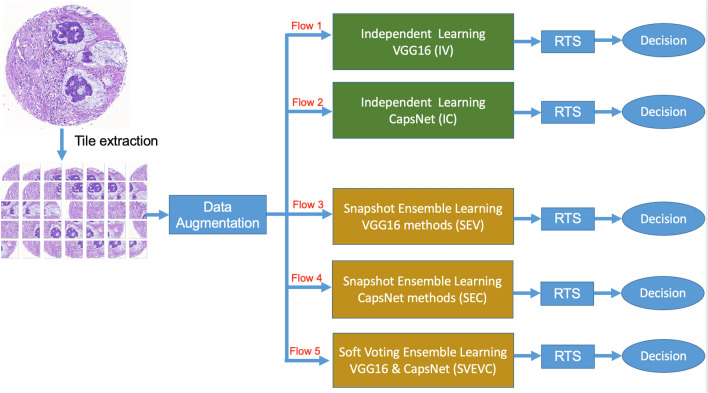
Tissue microarray (TMA) core images are a treasure trove for artificial intelligence applications. However, a common problem of TMAs is multiple sectioning, which can change the content of the intended tissue core and requires re-labelling. Here, we investigate different ensemble methods for colorectal tissue classification using high-throughput TMAs. Hematoxylin and Eosin (H&E) core images of 0.6 mm or 1.0 mm diameter from three international cohorts were extracted from 54 digital slides (n = 15,150 cores). After TMA core extraction and color enhancement, five different flows of independent and ensemble deep learning were applied. Training and testing data with 2144 and 13,006 cores included three classes: tumor, normal or "other" tissue. Ground-truth data were collected from 30 ngTMA slides (n = 8689 cores). A test augmentation is applied to reduce the uncertain prediction. Predictive accuracy of the best method, namely Soft Voting Ensemble of one VGG and one CapsNet models was 0.982, 0.947 and 0.939 for normal, "other" and tumor, which outperformed to independent or ensemble learning with one base-estimator. Our high-accuracy algorithm for colorectal tissue classification in high-throughput TMAs is amenable to images from different institutions, core sizes and stain intensity. It helps to reduce error in TMA core evaluations with previously given labels.

摘要

组织微阵列(TMA)核心图像是人工智能应用的宝库。然而,TMA 的一个常见问题是多次切片,这可能会改变预期组织核心的内容,并需要重新标记。在这里,我们研究了使用高通量 TMA 进行结直肠组织分类的不同集成方法。从三个国际队列的 54 张数字幻灯片中提取了直径为 0.6mm 或 1.0mm 的苏木精和伊红(H&E)核心图像(n=15150 个核心)。在 TMA 核心提取和颜色增强后,应用了五种不同的独立和集成深度学习流。包含三个类别的训练和测试数据:肿瘤、正常或“其他”组织,分别有 2144 个和 13006 个核心。从 30ngTMA 幻灯片(n=8689 个核心)收集了真实数据。应用测试扩充以减少不确定预测。最佳方法,即一个 VGG 和一个 CapsNet 模型的软投票集成的预测准确性分别为 0.982、0.947 和 0.939,用于正常、“其他”和肿瘤,优于具有一个基础估计器的独立或集成学习。我们用于高通量 TMA 中结直肠组织分类的高精度算法适用于来自不同机构、核心大小和染色强度的图像。它有助于减少具有给定标签的 TMA 核心评估中的错误。

https://cdn.ncbi.nlm.nih.gov/pmc/blobs/4419/7840737/7372c0036a62/41598_2021_81352_Fig6_HTML.jpg
https://cdn.ncbi.nlm.nih.gov/pmc/blobs/4419/7840737/0621a1db7ae6/41598_2021_81352_Fig1_HTML.jpg
https://cdn.ncbi.nlm.nih.gov/pmc/blobs/4419/7840737/db241d0d4959/41598_2021_81352_Fig2_HTML.jpg
https://cdn.ncbi.nlm.nih.gov/pmc/blobs/4419/7840737/8bf22fcf4957/41598_2021_81352_Fig3_HTML.jpg
https://cdn.ncbi.nlm.nih.gov/pmc/blobs/4419/7840737/ca31f9ef7292/41598_2021_81352_Fig4_HTML.jpg
https://cdn.ncbi.nlm.nih.gov/pmc/blobs/4419/7840737/96ec25ddb3c3/41598_2021_81352_Fig5_HTML.jpg
https://cdn.ncbi.nlm.nih.gov/pmc/blobs/4419/7840737/7372c0036a62/41598_2021_81352_Fig6_HTML.jpg

相似文献

1
Classification of colorectal tissue images from high throughput tissue microarrays by ensemble deep learning methods.基于集成深度学习方法的高通量组织微阵列中结直肠组织图像分类。
Sci Rep. 2021 Jan 27;11(1):2371. doi: 10.1038/s41598-021-81352-y.
2
Prognostic significance of mesothelin expression in colorectal cancer disclosed by area-specific four-point tissue microarrays.结直肠癌中特定区域四点组织微阵列检测间皮素表达的预后意义。
Virchows Arch. 2020 Sep;477(3):409-420. doi: 10.1007/s00428-020-02775-y. Epub 2020 Feb 27.
3
High throughput assessment of biomarkers in tissue microarrays using artificial intelligence: PTEN loss as a proof-of-principle in multi-center prostate cancer cohorts.利用人工智能对组织微阵列中的生物标志物进行高通量评估:多中心前列腺癌队列中 PTEN 缺失作为原理验证。
Mod Pathol. 2021 Feb;34(2):478-489. doi: 10.1038/s41379-020-00674-w. Epub 2020 Sep 3.
4
A next-generation tissue microarray (ngTMA) protocol for biomarker studies.一种用于生物标志物研究的新一代组织微阵列(ngTMA)方案。
J Vis Exp. 2014 Sep 23(91):51893. doi: 10.3791/51893.
5
Deep learning-based image analysis methods for brightfield-acquired multiplex immunohistochemistry images.基于深度学习的明场获取多重免疫组化图像的图像分析方法。
Diagn Pathol. 2020 Jul 28;15(1):100. doi: 10.1186/s13000-020-01003-0.
6
Next-generation tissue microarray (ngTMA) increases the quality of biomarker studies: an example using CD3, CD8, and CD45RO in the tumor microenvironment of six different solid tumor types.新一代组织微阵列(ngTMA)提高了生物标志物研究的质量:以六种不同实体瘤类型的肿瘤微环境中CD3、CD8和CD45RO为例。
J Transl Med. 2013 Apr 30;11:104. doi: 10.1186/1479-5876-11-104.
7
A TMA de-arraying method for high throughput biomarker discovery in tissue research.一种用于组织研究中高通量生物标志物发现的 TMA 解阵列方法。
PLoS One. 2011;6(10):e26007. doi: 10.1371/journal.pone.0026007. Epub 2011 Oct 7.
8
Robust gridding of TMAs after whole-slide imaging using template matching.使用模板匹配对全切片成像后的 TMA 进行稳健的网格化。
Cytometry A. 2010 Dec;77(12):1169-76. doi: 10.1002/cyto.a.20949.
9
Use and validation of epithelial recognition and fields of view algorithms on virtual slides to guide TMA construction.上皮识别和视野算法在虚拟切片上的应用及验证以指导组织微阵列构建。
Biotechniques. 2009 Nov;47(5):927-38. doi: 10.2144/000113207.
10
Automated curation of large-scale cancer histopathology image datasets using deep learning.利用深度学习对大规模癌症组织病理学图像数据集进行自动化注释。
Histopathology. 2024 Jun;84(7):1139-1153. doi: 10.1111/his.15159. Epub 2024 Feb 26.

引用本文的文献

1
Novel immunotherapeutics against LGR5 to target multiple cancer types.针对多种癌症类型的新型 LGR5 免疫疗法。
EMBO Mol Med. 2024 Sep;16(9):2233-2261. doi: 10.1038/s44321-024-00121-2. Epub 2024 Aug 21.
2
Topographic analysis of pancreatic cancer by TMA and digital spatial profiling reveals biological complexity with potential therapeutic implications.基于 TMA 和数字空间分析的胰腺癌的地形分析揭示了具有潜在治疗意义的生物学复杂性。
Sci Rep. 2024 May 18;14(1):11361. doi: 10.1038/s41598-024-62031-0.
3
3D multiplexed tissue imaging reconstruction and optimized region of interest (ROI) selection through deep learning model of channels embedding.

本文引用的文献

1
Explainable classifier for improving the accountability in decision-making for colorectal cancer diagnosis from histopathological images.用于提高基于组织病理学图像的结直肠癌诊断决策可解释性的分类器。
J Biomed Inform. 2020 Sep;109:103523. doi: 10.1016/j.jbi.2020.103523. Epub 2020 Aug 3.
2
Image-based consensus molecular subtype (imCMS) classification of colorectal cancer using deep learning.基于图像的共识分子亚型(imCMS)分类用于结直肠癌的深度学习。
Gut. 2021 Mar;70(3):544-554. doi: 10.1136/gutjnl-2019-319866. Epub 2020 Jul 20.
3
Artificial Intelligence Algorithms to Assess Hormonal Status From Tissue Microarrays in Patients With Breast Cancer.
通过通道嵌入深度学习模型进行3D多重组织成像重建及优化感兴趣区域(ROI)选择
Front Bioinform. 2023 Oct 19;3:1275402. doi: 10.3389/fbinf.2023.1275402. eCollection 2023.
4
TMA-Mate: An open-source modular toolkit for constructing tissue microarrays of arbitrary layouts.TMA-Mate:一种用于构建任意布局组织微阵列的开源模块化工具包。
HardwareX. 2023 Apr 11;14:e00419. doi: 10.1016/j.ohx.2023.e00419. eCollection 2023 Jun.
5
Deep learning-based image analysis predicts PD-L1 status from H&E-stained histopathology images in breast cancer.基于深度学习的图像分析可从乳腺癌的 H&E 染色组织病理学图像预测 PD-L1 状态。
Nat Commun. 2022 Nov 8;13(1):6753. doi: 10.1038/s41467-022-34275-9.
6
Deep Learning on Histopathological Images for Colorectal Cancer Diagnosis: A Systematic Review.用于结直肠癌诊断的组织病理学图像深度学习:一项系统综述。
Diagnostics (Basel). 2022 Mar 29;12(4):837. doi: 10.3390/diagnostics12040837.
7
COVID-CCD-Net: COVID-19 and colon cancer diagnosis system with optimized CNN hyperparameters using gradient-based optimizer.COVID-CCD-Net:基于梯度优化器优化 CNN 超参数的 COVID-19 和结肠癌诊断系统。
Med Biol Eng Comput. 2022 Jun;60(6):1595-1612. doi: 10.1007/s11517-022-02553-9. Epub 2022 Apr 8.
人工智能算法评估乳腺癌患者组织微阵列中的激素状态。
JAMA Netw Open. 2019 Jul 3;2(7):e197700. doi: 10.1001/jamanetworkopen.2019.7700.
4
Cell Line Derived Xenograft Mouse Models Are a Suitable Model for Studying Tumor Budding in Colorectal Cancer.细胞系衍生的异种移植小鼠模型是研究结直肠癌肿瘤芽生的合适模型。
Front Med (Lausanne). 2019 Jun 27;6:139. doi: 10.3389/fmed.2019.00139. eCollection 2019.
5
Deep learning can predict microsatellite instability directly from histology in gastrointestinal cancer.深度学习可直接从胃肠道癌症的组织学预测微卫星不稳定性。
Nat Med. 2019 Jul;25(7):1054-1056. doi: 10.1038/s41591-019-0462-y. Epub 2019 Jun 3.
6
Breast cancer outcome prediction with tumour tissue images and machine learning.利用肿瘤组织图像和机器学习预测乳腺癌的预后。
Breast Cancer Res Treat. 2019 Aug;177(1):41-52. doi: 10.1007/s10549-019-05281-1. Epub 2019 May 22.
7
Deep Learning With Sampling in Colon Cancer Histology.结肠癌组织学中带采样的深度学习
Front Bioeng Biotechnol. 2019 Mar 27;7:52. doi: 10.3389/fbioe.2019.00052. eCollection 2019.
8
Predicting survival from colorectal cancer histology slides using deep learning: A retrospective multicenter study.利用深度学习预测结直肠癌组织学切片的生存情况:一项回顾性多中心研究。
PLoS Med. 2019 Jan 24;16(1):e1002730. doi: 10.1371/journal.pmed.1002730. eCollection 2019 Jan.
9
MILD-Net: Minimal information loss dilated network for gland instance segmentation in colon histology images.MILD-Net:用于结肠组织学图像中腺体实例分割的最小信息丢失扩张网络。
Med Image Anal. 2019 Feb;52:199-211. doi: 10.1016/j.media.2018.12.001. Epub 2018 Dec 20.
10
Automated Gleason grading of prostate cancer tissue microarrays via deep learning.基于深度学习的前列腺癌组织微阵列 Gleason 分级自动化。
Sci Rep. 2018 Aug 13;8(1):12054. doi: 10.1038/s41598-018-30535-1.