• 文献检索
  • 文档翻译
  • 深度研究
  • 学术资讯
  • Suppr Zotero 插件Zotero 插件
  • 邀请有礼
  • 套餐&价格
  • 历史记录
应用&插件
Suppr Zotero 插件Zotero 插件浏览器插件Mac 客户端Windows 客户端微信小程序
定价
高级版会员购买积分包购买API积分包
服务
文献检索文档翻译深度研究API 文档MCP 服务
关于我们
关于 Suppr公司介绍联系我们用户协议隐私条款
关注我们

Suppr 超能文献

核心技术专利:CN118964589B侵权必究
粤ICP备2023148730 号-1Suppr @ 2026

文献检索

告别复杂PubMed语法,用中文像聊天一样搜索,搜遍4000万医学文献。AI智能推荐,让科研检索更轻松。

立即免费搜索

文件翻译

保留排版,准确专业,支持PDF/Word/PPT等文件格式,支持 12+语言互译。

免费翻译文档

深度研究

AI帮你快速写综述,25分钟生成高质量综述,智能提取关键信息,辅助科研写作。

立即免费体验

静态和运动细胞模型中的点、条纹和螺旋波:细胞中的 GTPase 模式。

Spots, stripes, and spiral waves in models for static and motile cells : GTPase patterns in cells.

机构信息

Department of Mathematics, University of British Columbia, Vancouver, V6T 1Z2, BC, Canada.

Mathematical Institute, University of Oxford, Oxford, OX2 6GG, UK.

出版信息

J Math Biol. 2021 Mar 4;82(4):28. doi: 10.1007/s00285-021-01550-0.

DOI:10.1007/s00285-021-01550-0
PMID:33660145
原文链接:https://pmc.ncbi.nlm.nih.gov/articles/PMC7929972/
Abstract

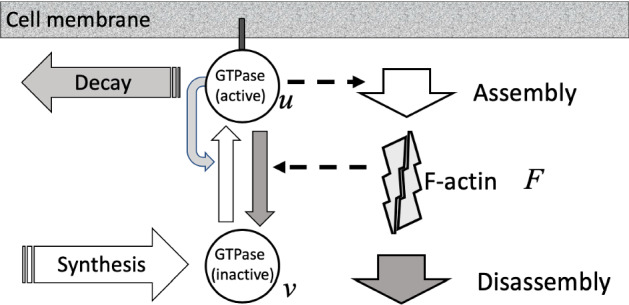
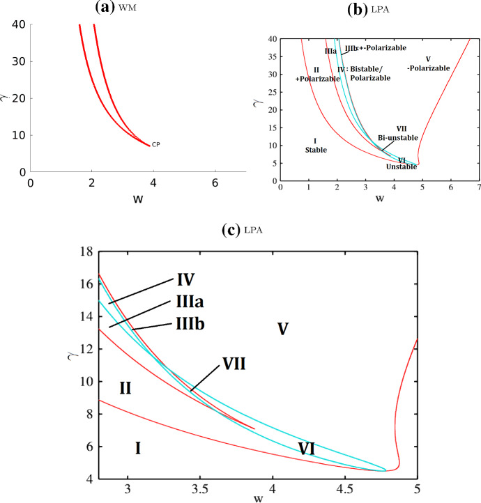
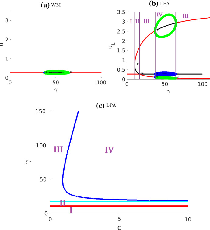
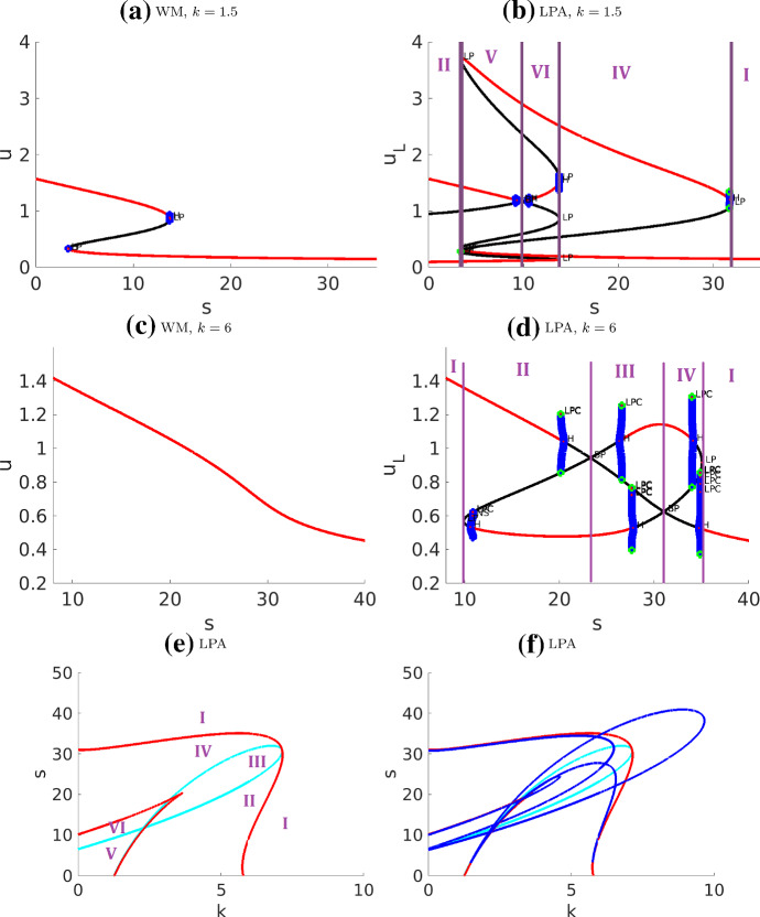
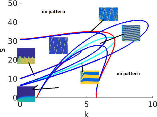
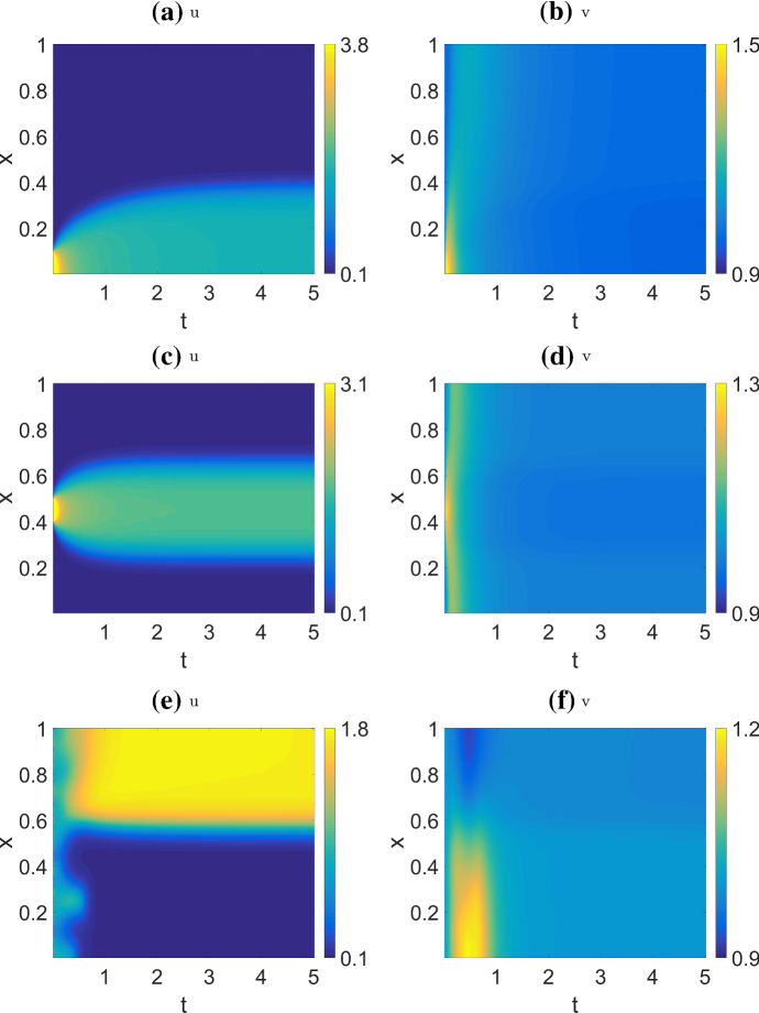
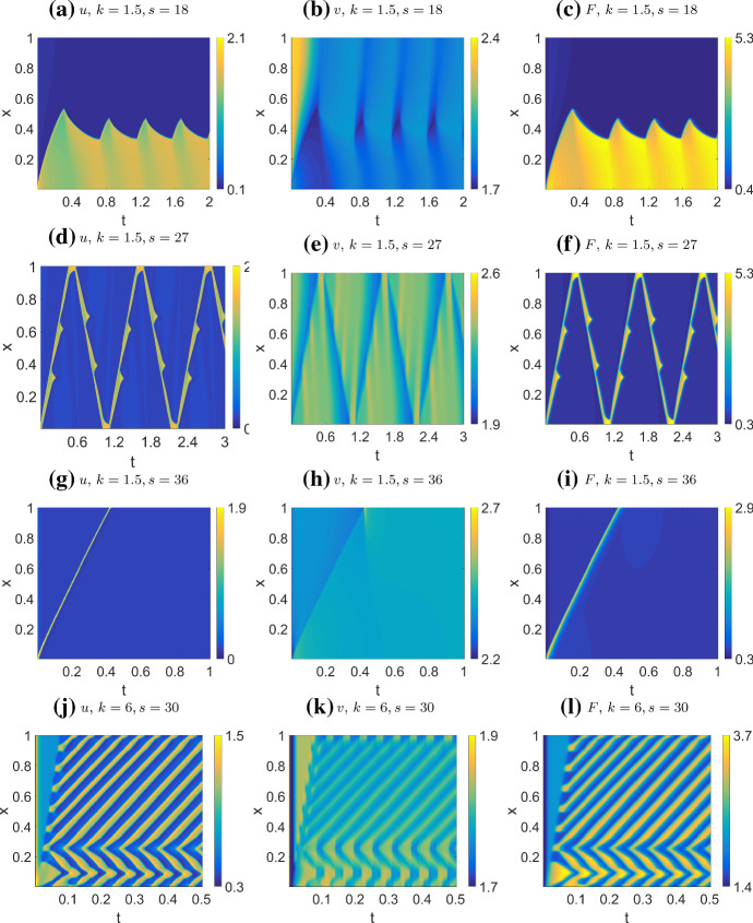
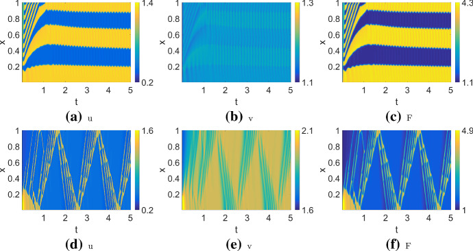
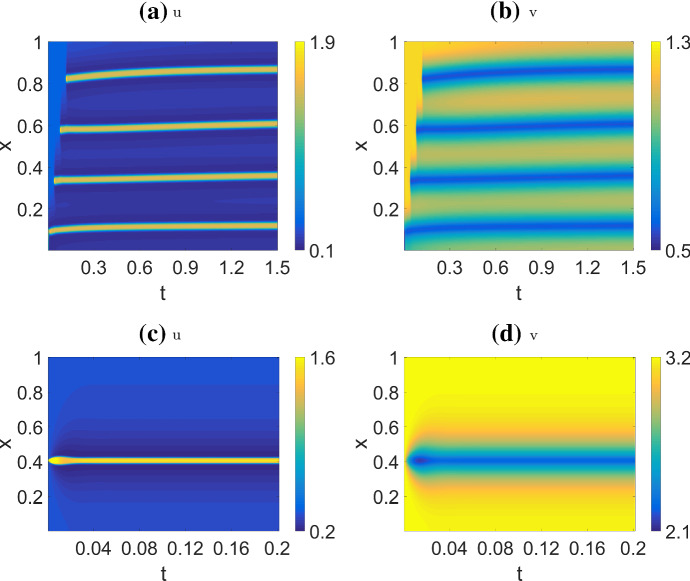
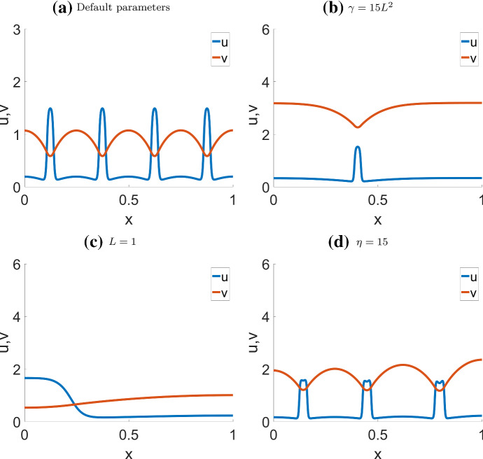
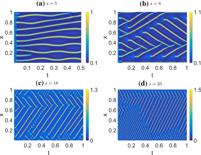
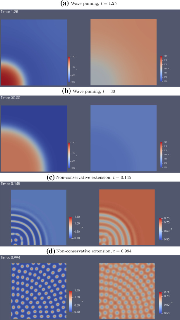
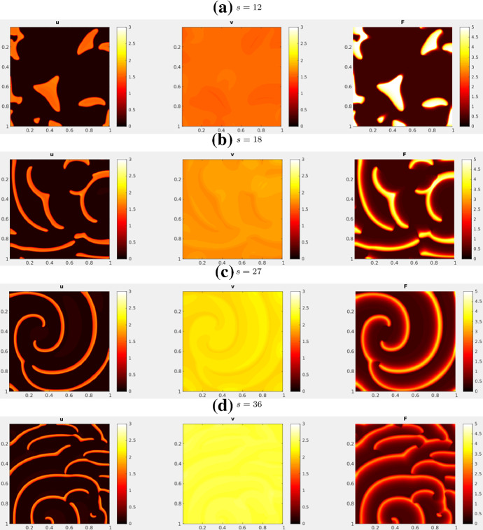
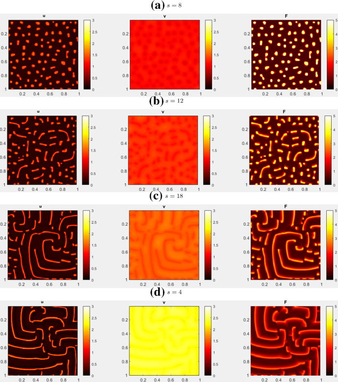
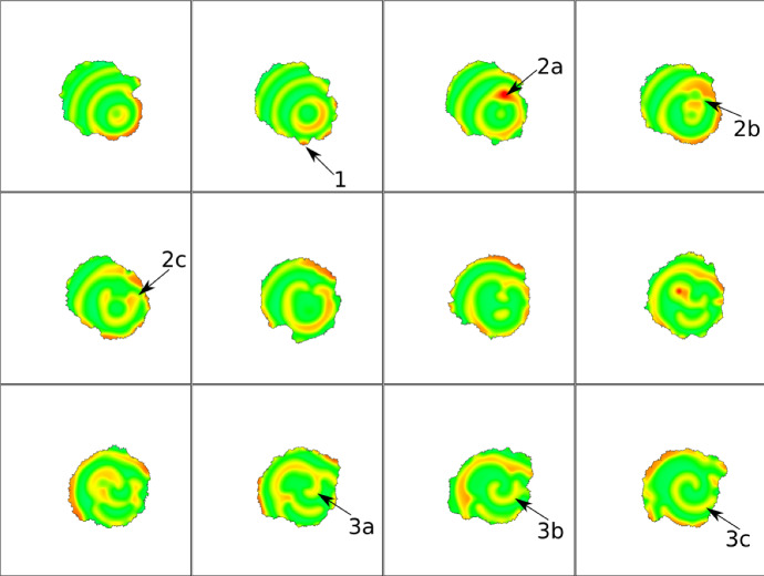
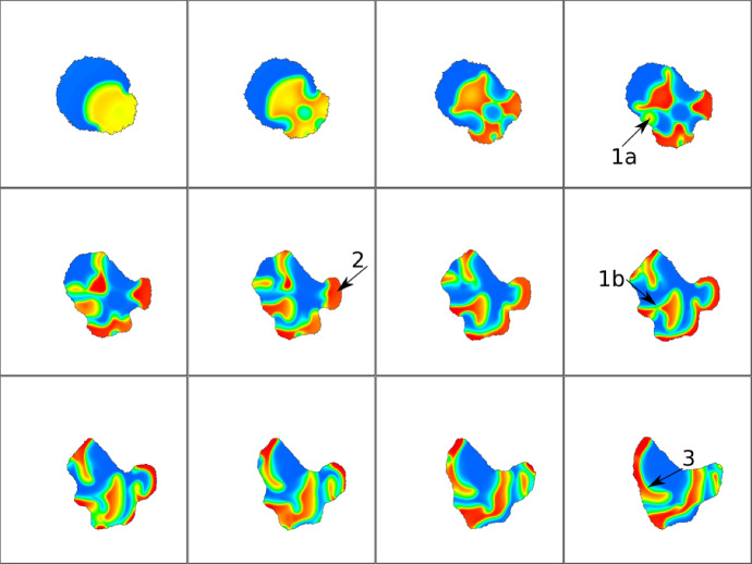
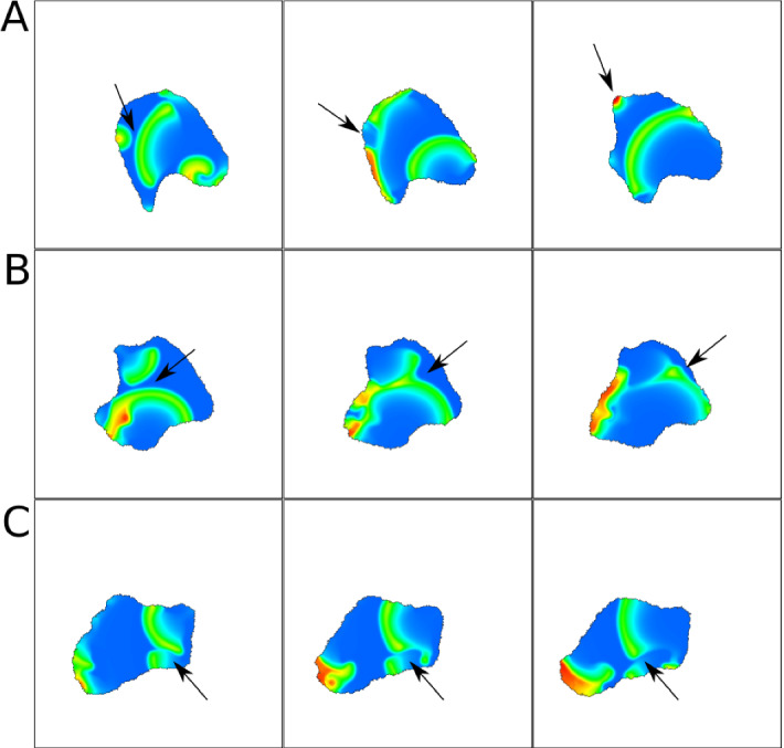
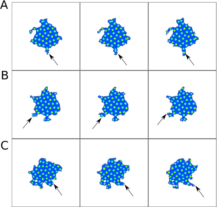
The polarization and motility of eukaryotic cells depends on assembly and contraction of the actin cytoskeleton and its regulation by proteins called GTPases. The activity of GTPases causes assembly of filamentous actin (by GTPases Cdc42, Rac), resulting in protrusion of the cell edge. Mathematical models for GTPase dynamics address the spontaneous formation of patterns and nonuniform spatial distributions of such proteins in the cell. Here we revisit the wave-pinning model for GTPase-induced cell polarization, together with a number of extensions proposed in the literature. These include introduction of sources and sinks of active and inactive GTPase (by the group of A. Champneys), and negative feedback from F-actin to GTPase activity. We discuss these extensions singly and in combination, in 1D, and 2D static domains. We then show how the patterns that form (spots, waves, and spirals) interact with cell boundaries to create a variety of interesting and dynamic cell shapes and motion.

摘要

真核细胞的极化和运动取决于肌动蛋白细胞骨架的组装和收缩,以及其调节蛋白 GTP 酶的作用。GTP 酶的活性导致丝状肌动蛋白的组装(由 GTP 酶 Cdc42、Rac 引起),从而导致细胞边缘的突出。用于 GTP 酶动力学的数学模型解决了此类蛋白质在细胞中自发形成模式和非均匀空间分布的问题。在这里,我们重新审视了 GTP 酶诱导的细胞极化的波钉模型,以及文献中提出的一些扩展。这些扩展包括引入活性和非活性 GTP 酶的源和汇(由 A. Champneys 小组提出),以及 F-肌动蛋白对 GTP 酶活性的负反馈。我们单独和组合地在 1D 和 2D 静态域中讨论了这些扩展。然后,我们展示了形成的模式(斑点、波和螺旋)如何与细胞边界相互作用,以创建各种有趣和动态的细胞形状和运动。

https://cdn.ncbi.nlm.nih.gov/pmc/blobs/bb84/7929972/2743837b4536/285_2021_1550_Fig24_HTML.jpg
https://cdn.ncbi.nlm.nih.gov/pmc/blobs/bb84/7929972/643ccc1456b5/285_2021_1550_Fig1_HTML.jpg
https://cdn.ncbi.nlm.nih.gov/pmc/blobs/bb84/7929972/79a1585f8e8d/285_2021_1550_Fig2_HTML.jpg
https://cdn.ncbi.nlm.nih.gov/pmc/blobs/bb84/7929972/619f35f8635f/285_2021_1550_Fig3_HTML.jpg
https://cdn.ncbi.nlm.nih.gov/pmc/blobs/bb84/7929972/c9a1f5fbc797/285_2021_1550_Fig4_HTML.jpg
https://cdn.ncbi.nlm.nih.gov/pmc/blobs/bb84/7929972/0d8c43e31d99/285_2021_1550_Fig5_HTML.jpg
https://cdn.ncbi.nlm.nih.gov/pmc/blobs/bb84/7929972/864bf97e390f/285_2021_1550_Fig6_HTML.jpg
https://cdn.ncbi.nlm.nih.gov/pmc/blobs/bb84/7929972/6bce75cd9d1e/285_2021_1550_Fig7_HTML.jpg
https://cdn.ncbi.nlm.nih.gov/pmc/blobs/bb84/7929972/4907638b41ef/285_2021_1550_Fig8_HTML.jpg
https://cdn.ncbi.nlm.nih.gov/pmc/blobs/bb84/7929972/8c92afaa92a5/285_2021_1550_Fig9_HTML.jpg
https://cdn.ncbi.nlm.nih.gov/pmc/blobs/bb84/7929972/2c3a1dbd38f9/285_2021_1550_Fig10_HTML.jpg
https://cdn.ncbi.nlm.nih.gov/pmc/blobs/bb84/7929972/212ae0cb1e8e/285_2021_1550_Fig11_HTML.jpg
https://cdn.ncbi.nlm.nih.gov/pmc/blobs/bb84/7929972/dc37ea703794/285_2021_1550_Fig12_HTML.jpg
https://cdn.ncbi.nlm.nih.gov/pmc/blobs/bb84/7929972/83dee3c1c72d/285_2021_1550_Fig13_HTML.jpg
https://cdn.ncbi.nlm.nih.gov/pmc/blobs/bb84/7929972/eb4ff17a18b4/285_2021_1550_Fig14_HTML.jpg
https://cdn.ncbi.nlm.nih.gov/pmc/blobs/bb84/7929972/381ffdc57db5/285_2021_1550_Fig15_HTML.jpg
https://cdn.ncbi.nlm.nih.gov/pmc/blobs/bb84/7929972/31d9bc3012f1/285_2021_1550_Fig16_HTML.jpg
https://cdn.ncbi.nlm.nih.gov/pmc/blobs/bb84/7929972/b249ec7a1427/285_2021_1550_Fig17_HTML.jpg
https://cdn.ncbi.nlm.nih.gov/pmc/blobs/bb84/7929972/94468ec9190a/285_2021_1550_Fig18_HTML.jpg
https://cdn.ncbi.nlm.nih.gov/pmc/blobs/bb84/7929972/d8ef3e3c308c/285_2021_1550_Fig19_HTML.jpg
https://cdn.ncbi.nlm.nih.gov/pmc/blobs/bb84/7929972/527f1382938f/285_2021_1550_Fig20_HTML.jpg
https://cdn.ncbi.nlm.nih.gov/pmc/blobs/bb84/7929972/417bdf0d10c8/285_2021_1550_Fig21_HTML.jpg
https://cdn.ncbi.nlm.nih.gov/pmc/blobs/bb84/7929972/5c6c48cbfafa/285_2021_1550_Fig22_HTML.jpg
https://cdn.ncbi.nlm.nih.gov/pmc/blobs/bb84/7929972/d597043fc92e/285_2021_1550_Fig23_HTML.jpg
https://cdn.ncbi.nlm.nih.gov/pmc/blobs/bb84/7929972/2743837b4536/285_2021_1550_Fig24_HTML.jpg

相似文献

1
Spots, stripes, and spiral waves in models for static and motile cells : GTPase patterns in cells.静态和运动细胞模型中的点、条纹和螺旋波:细胞中的 GTPase 模式。
J Math Biol. 2021 Mar 4;82(4):28. doi: 10.1007/s00285-021-01550-0.
2
Cell Size, Mechanical Tension, and GTPase Signaling in the Single Cell.单细胞中的细胞大小、机械张力和 GTPase 信号转导。
Bull Math Biol. 2020 Feb 3;82(2):28. doi: 10.1007/s11538-020-00702-5.
3
Regimes of wave type patterning driven by refractory actin feedback: transition from static polarization to dynamic wave behaviour.由反射性肌动蛋白反馈驱动的波型模式的时相:从静态极化到动态波行为的转变。
Phys Biol. 2012 Aug;9(4):046005. doi: 10.1088/1478-3975/9/4/046005. Epub 2012 Jul 11.
4
Visualization and molecular analysis of actin assembly in living cells.活细胞中肌动蛋白组装的可视化与分子分析
J Cell Biol. 1998 Dec 28;143(7):1919-30. doi: 10.1083/jcb.143.7.1919.
5
Ras-related GTPases and the cytoskeleton.Ras相关的GTP酶与细胞骨架。
Mol Biol Cell. 1992 May;3(5):475-9. doi: 10.1091/mbc.3.5.475.
6
Analysis of a minimal Rho-GTPase circuit regulating cell shape.调控细胞形状的最小Rho-GTPase信号通路分析
Phys Biol. 2016 Jul 19;13(4):046001. doi: 10.1088/1478-3975/13/4/046001.
7
Actin dynamics in cell migration.肌动蛋白在细胞迁移中的动态变化。
Essays Biochem. 2019 Oct 31;63(5):483-495. doi: 10.1042/EBC20190015.
8
Role of actin polymerization and adhesion to extracellular matrix in Rac- and Rho-induced cytoskeletal reorganization.肌动蛋白聚合作用及与细胞外基质的黏附在Rac和Rho诱导的细胞骨架重组中的作用
J Cell Biol. 1997 Aug 25;138(4):913-26. doi: 10.1083/jcb.138.4.913.
9
The role of Rho GTPases in the regulation of the rearrangement of actin cytoskeleton and cell movement.Rho GTP 酶在肌动蛋白细胞骨架重排和细胞运动调节中的作用。
Exp Mol Med. 2004 Aug 31;36(4):358-66. doi: 10.1038/emm.2004.47.
10
A mechanochemical model for rho GTPase mediated cell polarization.一种用于Rho GTP酶介导的细胞极化的机械化学模型。
J Theor Biol. 2020 Nov 7;504:110386. doi: 10.1016/j.jtbi.2020.110386. Epub 2020 Jul 9.

引用本文的文献

1
Investigating local negative feedback of Rac activity by mathematical models and cell motility simulations.通过数学模型和细胞运动模拟研究Rac活性的局部负反馈。
bioRxiv. 2025 May 5:2025.05.05.651928. doi: 10.1101/2025.05.05.651928.
2
Rho of Plants patterning: linking mathematical models and molecular diversity.植物形态形成中的 Rho 蛋白:连接数学模型和分子多样性。
J Exp Bot. 2024 Feb 28;75(5):1274-1288. doi: 10.1093/jxb/erad447.
3
From actin waves to mechanism and back: How theory aids biological understanding.从肌动蛋白波到机制再到理论:理论如何帮助生物理解。

本文引用的文献

1
Cell Size, Mechanical Tension, and GTPase Signaling in the Single Cell.单细胞中的细胞大小、机械张力和 GTPase 信号转导。
Bull Math Biol. 2020 Feb 3;82(2):28. doi: 10.1007/s11538-020-00702-5.
2
Excitable RhoA dynamics drive pulsed contractions in the early embryo.激动的 RhoA 动力学驱动早期胚胎的脉冲收缩。
J Cell Biol. 2018 Dec 3;217(12):4230-4252. doi: 10.1083/jcb.201806161. Epub 2018 Oct 1.
3
Coupling mechanical tension and GTPase signaling to generate cell and tissue dynamics.耦合机械张力与小GTP酶信号传导以产生细胞和组织动力学。
Elife. 2023 Jul 10;12:e87181. doi: 10.7554/eLife.87181.
4
Spatiotemporal Coordination of Rac1 and Cdc42 at the Whole Cell Level during Cell Ruffling.细胞皱襞过程中 Rac1 和 Cdc42 在全细胞水平上的时空协调
Cells. 2023 Jun 15;12(12):1638. doi: 10.3390/cells12121638.
5
Dynamic regulation of RAS and RAS signaling.RAS 及其信号通路的动态调控。
Biochem J. 2023 Jan 13;480(1):1-23. doi: 10.1042/BCJ20220234.
6
Spatial models of pattern formation during phagocytosis.吞噬作用过程中模式形成的空间模型。
PLoS Comput Biol. 2022 Oct 3;18(10):e1010092. doi: 10.1371/journal.pcbi.1010092. eCollection 2022 Oct.
7
Nonmodular oscillator and switch based on RNA decay drive regeneration of multimodal gene expression.基于 RNA 衰减的非模块化振荡器和开关驱动多模态基因表达的再生。
Nucleic Acids Res. 2022 Apr 22;50(7):3693-3708. doi: 10.1093/nar/gkac217.
8
Modern perspectives on near-equilibrium analysis of Turing systems.现代视角下的图灵系统近平衡分析
Philos Trans A Math Phys Eng Sci. 2021 Dec 27;379(2213):20200268. doi: 10.1098/rsta.2020.0268. Epub 2021 Nov 8.
9
Spatiotemporal development of coexisting wave domains of Rho activity in the cell cortex.细胞皮层中 Rho 活性共存波域的时空发展。
Sci Rep. 2021 Sep 30;11(1):19512. doi: 10.1038/s41598-021-99029-x.
10
From the Matrix to the Nucleus and Back: Mechanobiology in the Light of Health, Pathologies, and Regeneration of Oral Periodontal Tissues.从基质到细胞核再回来:口腔牙周组织的健康、病理和再生中的机械生物学。
Biomolecules. 2021 May 31;11(6):824. doi: 10.3390/biom11060824.
Phys Biol. 2018 Apr 30;15(4):046004. doi: 10.1088/1478-3975/aab1c0.
4
Mechanisms of Cell Polarization.细胞极化的机制
Curr Opin Syst Biol. 2017 Jun;3:43-53. doi: 10.1016/j.coisb.2017.03.005. Epub 2017 Apr 12.
5
Actin Waves: Origin of Cell Polarization and Migration?肌动蛋白波:细胞极化和迁移的起源?
Trends Cell Biol. 2017 Jul;27(7):515-526. doi: 10.1016/j.tcb.2017.02.003. Epub 2017 Mar 7.
6
Analysis of a minimal Rho-GTPase circuit regulating cell shape.调控细胞形状的最小Rho-GTPase信号通路分析
Phys Biol. 2016 Jul 19;13(4):046001. doi: 10.1088/1478-3975/13/4/046001.
7
The biophysical nature of cells: potential cell behaviours revealed by analytical and computational studies of cell surface mechanics.细胞的生物物理特性:通过细胞表面力学的分析和计算研究揭示的潜在细胞行为。
BMC Biophys. 2015 May 12;8:8. doi: 10.1186/s13628-015-0022-x. eCollection 2015.
8
Local perturbation analysis: a computational tool for biophysical reaction-diffusion models.局部微扰分析:一种用于生物物理反应扩散模型的计算工具。
Biophys J. 2015 Jan 20;108(2):230-6. doi: 10.1016/j.bpj.2014.11.3457.
9
An efficient, nonlinear stability analysis for detecting pattern formation in reaction diffusion systems.一种用于检测反应扩散系统中模式形成的高效非线性稳定性分析方法。
Bull Math Biol. 2014 Jan;76(1):157-83. doi: 10.1007/s11538-013-9914-6. Epub 2013 Oct 25.
10
From simple to detailed models for cell polarization.从简单到详细的细胞极化模型。
Philos Trans R Soc Lond B Biol Sci. 2013 Sep 23;368(1629):20130003. doi: 10.1098/rstb.2013.0003. Print 2013.