• 文献检索
  • 文档翻译
  • 深度研究
  • 学术资讯
  • Suppr Zotero 插件Zotero 插件
  • 邀请有礼
  • 套餐&价格
  • 历史记录
应用&插件
Suppr Zotero 插件Zotero 插件浏览器插件Mac 客户端Windows 客户端微信小程序
定价
高级版会员购买积分包购买API积分包
服务
文献检索文档翻译深度研究API 文档MCP 服务
关于我们
关于 Suppr公司介绍联系我们用户协议隐私条款
关注我们

Suppr 超能文献

核心技术专利:CN118964589B侵权必究
粤ICP备2023148730 号-1Suppr @ 2026

文献检索

告别复杂PubMed语法,用中文像聊天一样搜索,搜遍4000万医学文献。AI智能推荐,让科研检索更轻松。

立即免费搜索

文件翻译

保留排版,准确专业,支持PDF/Word/PPT等文件格式,支持 12+语言互译。

免费翻译文档

深度研究

AI帮你快速写综述,25分钟生成高质量综述,智能提取关键信息,辅助科研写作。

立即免费体验

2019冠状病毒病(COVID-19)肺炎胸部CT分级系统的比较

Comparison of Chest CT Grading Systems in Coronavirus Disease 2019 (COVID-19) Pneumonia.

作者信息

Inui Shohei, Kurokawa Ryo, Nakai Yudai, Watanabe Yusuke, Kurokawa Mariko, Sakurai Keita, Fujikawa Akira, Sugiura Hiroaki, Kawahara Takuya, Yoon Soon Ho, Uwabe Yasuhide, Uchida Yuto, Gonoi Wataru, Abe Osamu

机构信息

Department of Radiology, Graduate School of Medicine, The University of Tokyo, 7-3-1, Hongo, Bunkyo-ku, Tokyo, 113-8655, Japan (S.I., R.K., Y.N., Y.W., W.G., O.A,); Department of Radiology, Japan Self-Defense Forces Central Hospital, 1-2-24, Ikejiri, Setagaya-ku, Tokyo, 154-0001, Japan (S.I., A.F.); Department of Radiology, Tokyo Metropolitan Cancer and Infectious Diseases Center Komagome Hospital, 3-18-22, Honkomagome, Bunkyo-ku, Tokyo, 113-8677, Japan (M.K.); Department of Radiology, National Center for Geriatrics and Gerontology, 7-430, Morioka-cho, Obu, Aichi, 474-8511, Japan (K.S.); Department of Radiology, National Defense Medical College, 3-2, Namiki, Tokorozawa-shi, Saitama, 359-8513, Japan (H.S.); Clinical Research Promotion Center, The University of Tokyo Hospital, 7-3-1, Hongo, Bunkyo-ku, Tokyo, 113-8655, Japan (T.K.); Department of Radiology, Seoul National University College of Medicine, Seoul National University Hospital, 101 Daehak-ro, Chongno-gu, Seoul 03080, Republic of Korea (S.H.Y.); Department of Respiratory Medicine, Japan Self-Defense Forces Central Hospital, 1-2-24, Ikejiri, Setagaya-ku, Tokyo, 154-0001, Japan (Y.U.); Department of Neurology and Neuroscience, Nagoya City University Graduate School of Medical Sciences, 1, Kawasumi, Mizuho-ku, Nagoya, 467-8601, Japan (Y.U.).

出版信息

Radiol Cardiothorac Imaging. 2020 Nov 5;2(6):e200492. doi: 10.1148/ryct.2020200492. eCollection 2020 Dec.

DOI:10.1148/ryct.2020200492
PMID:33778648
原文链接:https://pmc.ncbi.nlm.nih.gov/articles/PMC7650298/
Abstract

PURPOSE

To compare the performance and interobserver agreement of the COVID-19 Reporting and Data System (CO-RADS), the COVID-19 imaging reporting and data system (COVID-RADS), the RSNA expert consensus statement, and the British Society of Thoracic Imaging (BSTI) guidance statement.

MATERIALS AND METHODS

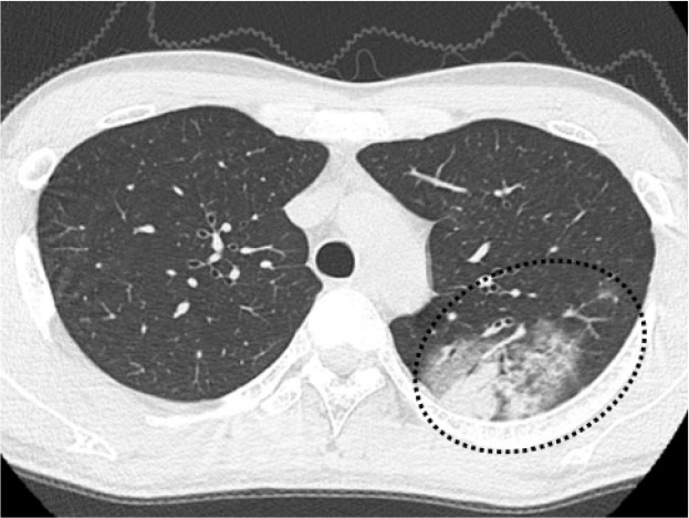
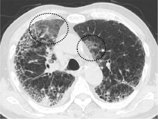
In this case-control study, total of 100 symptomatic patients suspected of having COVID-19 were included: 50 patients with COVID-19 (59±17 years, 38 men) and 50 patients without COVID-19 (65±24 years, 30 men). Eight radiologists independently scored chest CT images of the cohort according to each reporting system. The area under the receiver operating characteristic curves (AUC) and interobserver agreements were calculated and statistically compared across the systems.

RESULTS

A total of 800 observations were made for each system. The level of suspicion of COVID-19 correlated with the RT-PCR positive rate except for the "negative for pneumonia" classifications in all the systems (Spearman's coefficient: ρ=1.0, =<.001 for all the systems). Average AUCs were as follows: CO-RADS, 0.84 (95% confidence interval, 0.83-0.85): COVID-RADS, 0.80 (0.78-0.81): the RSNA statement, 0.81 (0.79-0.82): and the BSTI statement, 0.84 (0.812-0.86). Average Cohen's kappa across observers was 0.62 (95% confidence interval, 0.58-0.66), 0.63 (0.58-0.68), 0.63 (0.57-0.69), and 0.61 (0.58-0.64) for CO-RADS, COVID-RADS, the RSNA statement and the BSTI statement, respectively. CO-RADS and the BSTI statement outperformed COVID-RADS and the RSNA statement in diagnostic performance (=.<.05 for all the comparison).

CONCLUSIONS

CO-RADS, COVID-RADS, the RSNA statement and the BSTI statement provided reasonable performances and interobserver agreements in reporting CT findings of COVID-19.

摘要

目的

比较新型冠状病毒肺炎报告与数据系统(CO-RADS)、新型冠状病毒肺炎影像报告和数据系统(COVID-RADS)、美国放射学会(RSNA)专家共识声明以及英国胸科影像学会(BSTI)指南声明的性能及观察者间的一致性。

材料与方法

在这项病例对照研究中,共纳入100例疑似新型冠状病毒肺炎的有症状患者:50例新型冠状病毒肺炎患者(59±17岁,38例男性)和50例非新型冠状病毒肺炎患者(65±24岁,30例男性)。8名放射科医生根据每个报告系统对该队列的胸部CT图像进行独立评分。计算各系统的受试者操作特征曲线下面积(AUC)和观察者间一致性,并进行统计学比较。

结果

每个系统共进行了800次观察。除所有系统中的“肺炎阴性”分类外,新型冠状病毒肺炎的怀疑程度与逆转录聚合酶链反应(RT-PCR)阳性率相关(斯皮尔曼系数:所有系统中ρ = 1.0,P <.001)。平均AUC如下:CO-RADS为0.84(95%置信区间,0.83 - 0.85);COVID-RADS为0.80(0.78 - 0.81);RSNA声明为0.81(0.79 - 0.82);BSTI声明为0.84(0.812 - 0.86)。CO-RADS、COVID-RADS、RSNA声明和BSTI声明观察者间的平均科恩kappa系数分别为0.62(95%置信区间,0.58 - 0.66)、0.63(0.58 - 0.68)、0.63(0.57 - 0.69)和0.61(0.58 - 0.64)。在诊断性能方面,CO-RADS和BSTI声明优于COVID-RADS和RSNA声明(所有比较中P <.05)。

结论

CO-RADS、COVID-RADS、RSNA声明和BSTI声明在报告新型冠状病毒肺炎的CT表现时具有合理的性能和观察者间一致性。

https://cdn.ncbi.nlm.nih.gov/pmc/blobs/d16f/7977812/5c330c4b9f17/ryct.2020200492.fig5b.jpg
https://cdn.ncbi.nlm.nih.gov/pmc/blobs/d16f/7977812/df29e335fb3c/ryct.2020200492.fig1.jpg
https://cdn.ncbi.nlm.nih.gov/pmc/blobs/d16f/7977812/3cc0b52278b8/ryct.2020200492.fig2a.jpg
https://cdn.ncbi.nlm.nih.gov/pmc/blobs/d16f/7977812/587acac14db5/ryct.2020200492.fig2b.jpg
https://cdn.ncbi.nlm.nih.gov/pmc/blobs/d16f/7977812/5ecb13192f4b/ryct.2020200492.fig2c.jpg
https://cdn.ncbi.nlm.nih.gov/pmc/blobs/d16f/7977812/e7537945ceba/ryct.2020200492.fig3a.jpg
https://cdn.ncbi.nlm.nih.gov/pmc/blobs/d16f/7977812/75973bb00ceb/ryct.2020200492.fig3b.jpg
https://cdn.ncbi.nlm.nih.gov/pmc/blobs/d16f/7977812/12a0d6c1f875/ryct.2020200492.fig3c.jpg
https://cdn.ncbi.nlm.nih.gov/pmc/blobs/d16f/7977812/e3cbf5c12cd4/ryct.2020200492.fig3d.jpg
https://cdn.ncbi.nlm.nih.gov/pmc/blobs/d16f/7977812/6c93519bcd46/ryct.2020200492.fig4a.jpg
https://cdn.ncbi.nlm.nih.gov/pmc/blobs/d16f/7977812/40116add4a90/ryct.2020200492.fig4b.jpg
https://cdn.ncbi.nlm.nih.gov/pmc/blobs/d16f/7977812/c461863a8516/ryct.2020200492.fig4c.jpg
https://cdn.ncbi.nlm.nih.gov/pmc/blobs/d16f/7977812/bcf7cae2e4b1/ryct.2020200492.fig5a.jpg
https://cdn.ncbi.nlm.nih.gov/pmc/blobs/d16f/7977812/5c330c4b9f17/ryct.2020200492.fig5b.jpg

相似文献

1
Comparison of Chest CT Grading Systems in Coronavirus Disease 2019 (COVID-19) Pneumonia.2019冠状病毒病(COVID-19)肺炎胸部CT分级系统的比较
Radiol Cardiothorac Imaging. 2020 Nov 5;2(6):e200492. doi: 10.1148/ryct.2020200492. eCollection 2020 Dec.
2
RSNA and BSTI grading systems of COVID-19 pneumonia: comparison of the diagnostic performance and interobserver agreement.RSNA 和 BSTI 新冠肺炎肺炎分级系统:诊断性能和观察者间一致性比较。
BMC Med Imaging. 2021 Oct 4;21(1):143. doi: 10.1186/s12880-021-00668-3.
3
Standardized reporting systems of chest computed tomography in a population with low coronavirus disease 2019 prevalence: A retrospective comparative study.2019冠状病毒病低流行人群胸部计算机断层扫描标准化报告系统:一项回顾性比较研究。
Heliyon. 2021 Aug;7(8):e07743. doi: 10.1016/j.heliyon.2021.e07743. Epub 2021 Aug 10.
4
Radiological Society of North America (RSNA) Expert Consensus Statement Related to Chest CT Findings in COVID-19 Versus CO-RADS: Comparison of Reporting System Performance Among Chest Radiologists and End-User Preference.北美放射学会(RSNA)关于 COVID-19 与 CO-RADS 胸部 CT 表现的专家共识声明:胸部放射科医生和最终用户偏好之间报告系统性能的比较。
Can Assoc Radiol J. 2021 Nov;72(4):806-813. doi: 10.1177/0846537120968919. Epub 2020 Nov 3.
5
CO-RADS: A Categorical CT Assessment Scheme for Patients Suspected of Having COVID-19-Definition and Evaluation.CO-RADS:一种用于疑似 COVID-19 患者的 CT 分类评估方案——定义和评估。
Radiology. 2020 Aug;296(2):E97-E104. doi: 10.1148/radiol.2020201473. Epub 2020 Apr 27.
6
Comparison of the CO-RADS and the RSNA chest CT classification system concerning sensitivity and reliability for the diagnosis of COVID-19 pneumonia.CO-RADS与RSNA胸部CT分类系统在诊断新型冠状病毒肺炎的敏感性和可靠性方面的比较。
Insights Imaging. 2021 Apr 28;12(1):55. doi: 10.1186/s13244-021-00998-4.
7
Evaluation of the Usefulness of CO-RADS for Chest CT in Patients Suspected of Having COVID-19.CO-RADS在疑似新型冠状病毒肺炎患者胸部CT中的应用价值评估
Diagnostics (Basel). 2020 Aug 19;10(9):608. doi: 10.3390/diagnostics10090608.
8
RSNA Expert Consensus Statement on Reporting Chest CT Findings Related to COVID-19: Interobserver Agreement Between Chest Radiologists.RSNA 专家关于报告与 COVID-19 相关的胸部 CT 结果的共识声明:胸部放射科医生之间的观察者间一致性。
Can Assoc Radiol J. 2021 Feb;72(1):159-166. doi: 10.1177/0846537120938328. Epub 2020 Jul 2.
9
Automated Assessment of COVID-19 Reporting and Data System and Chest CT Severity Scores in Patients Suspected of Having COVID-19 Using Artificial Intelligence.利用人工智能对疑似 COVID-19 患者的 COVID-19 报告和数据系统及胸部 CT 严重程度评分进行自动评估。
Radiology. 2021 Jan;298(1):E18-E28. doi: 10.1148/radiol.2020202439. Epub 2020 Jul 30.
10
Diagnostic Performance of Chest CT for SARS-CoV-2 Infection in Individuals with or without COVID-19 Symptoms.胸部 CT 对有或无症状 COVID-19 患者 SARS-CoV-2 感染的诊断性能。
Radiology. 2021 Jan;298(1):E30-E37. doi: 10.1148/radiol.2020202708. Epub 2020 Aug 10.

引用本文的文献

1
"KAIZEN" method realizing implementation of deep-learning models for COVID-19 CT diagnosis in real world hospitals.“KAIZEN”方法实现 COVID-19 CT 诊断深度学习模型在真实世界医院中的应用。
Sci Rep. 2024 Jan 19;14(1):1672. doi: 10.1038/s41598-024-52135-y.
2
Assessment of COVID-19 RT-PCR Positive Symptomatic Patients With Clinical, Hematological, and Radiological Parameters Among Three Groups: A Comparative Study.三组中COVID-19逆转录聚合酶链反应阳性有症状患者的临床、血液学和放射学参数评估:一项比较研究
Cureus. 2023 May 30;15(5):e39681. doi: 10.7759/cureus.39681. eCollection 2023 May.
3
Diagnostic performance of standardized typical CT findings for COVID-19: a systematic review and meta-analysis.

本文引用的文献

1
Diagnostic Accuracy of North America Expert Consensus Statement on Reporting CT Findings in Patients Suspected of Having COVID-19 Infection: An Italian Single-Center Experience.北美关于报告疑似新型冠状病毒肺炎感染患者CT检查结果的专家共识声明的诊断准确性:一项意大利单中心经验
Radiol Cardiothorac Imaging. 2020 Jul 23;2(4):e200312. doi: 10.1148/ryct.2020200312. eCollection 2020 Aug.
2
Radiological Society of North America Chest CT Classification System for Reporting COVID-19 Pneumonia: Interobserver Variability and Correlation with Reverse-Transcription Polymerase Chain Reaction.北美放射学会用于报告新型冠状病毒肺炎的胸部CT分类系统:观察者间差异及与逆转录聚合酶链反应的相关性
Radiol Cardiothorac Imaging. 2020 Jun 11;2(3):e200213. doi: 10.1148/ryct.2020200213. eCollection 2020 Jun.
3
COVID-19标准化典型CT表现的诊断性能:一项系统评价和荟萃分析
Insights Imaging. 2023 May 24;14(1):96. doi: 10.1186/s13244-023-01429-2.
4
COVID-19 in a group of children with asthma: presentation, severity, and outcome.一组哮喘儿童中的新型冠状病毒肺炎:临床表现、严重程度及转归
Am J Clin Exp Immunol. 2022 Dec 15;11(6):92-102. eCollection 2022.
5
Is the generalizability of a developed artificial intelligence algorithm for COVID-19 on chest CT sufficient for clinical use? Results from the International Consortium for COVID-19 Imaging AI (ICOVAI).开发用于 COVID-19 胸部 CT 的人工智能算法的可推广性是否足以用于临床?来自 COVID-19 成像人工智能国际联合会(ICOVAI)的结果。
Eur Radiol. 2023 Jun;33(6):4249-4258. doi: 10.1007/s00330-022-09303-3. Epub 2023 Jan 18.
6
COVID-19 pneumonia: lessons learned, challenges, and preparing for the future.新型冠状病毒肺炎:经验教训、挑战和未来准备。
Diagn Interv Radiol. 2022 Nov;28(6):576-585. doi: 10.5152/dir.2022.221881.
7
Investigation of the relationship of CO-RADS and CT patterns with laboratory parameters in COVID-19 patients and a new perspective on the total CT scoring system.探讨 COVID-19 患者的 CO-RADS 与 CT 表现与实验室参数的关系和对 CT 总分系统的新认识。
BMC Med Imaging. 2022 Jul 20;22(1):128. doi: 10.1186/s12880-022-00857-8.
8
Chest CT Findings in Hospitalized Patients with SARS-CoV-2: Delta versus Omicron Variants.住院 SARS-CoV-2 感染患者的胸部 CT 表现:Delta 变异株与奥密克戎变异株。
Radiology. 2023 Jan;306(1):252-260. doi: 10.1148/radiol.220676. Epub 2022 Jun 28.
9
Clinical significance of chest CT for the exclusion of COVID-19 in pre-admission screening: Is it worthwhile using chest CT with reverse-transcription polymerase chain reaction test?胸部 CT 对疑似 COVID-19 患者入院前筛查的临床意义:与逆转录聚合酶链反应检测联合使用胸部 CT 是否有价值?
Respir Investig. 2022 Jul;60(4):595-603. doi: 10.1016/j.resinv.2022.04.007. Epub 2022 May 8.
10
Diagnostic performance of CO-RADS for COVID-19: a systematic review and meta-analysis.CO-RADS 对 COVID-19 的诊断性能:系统评价和荟萃分析。
Eur Radiol. 2022 Jul;32(7):4414-4426. doi: 10.1007/s00330-022-08576-y. Epub 2022 Mar 29.
Radiological Society of North America Expert Consensus Document on Reporting Chest CT Findings Related to COVID-19: Endorsed by the Society of Thoracic Radiology, the American College of Radiology, and RSNA.北美放射学会关于报告与COVID-19相关的胸部CT检查结果的专家共识文件:得到了胸腔放射学会、美国放射学会和北美放射学会的认可。
Radiol Cardiothorac Imaging. 2020 Mar 25;2(2):e200152. doi: 10.1148/ryct.2020200152. eCollection 2020 Apr.
4
Chest CT Findings in Cases from the Cruise Ship with Coronavirus Disease (COVID-19).邮轮上新型冠状病毒肺炎(COVID-19)病例的胸部CT表现
Radiol Cardiothorac Imaging. 2020 Mar 17;2(2):e200110. doi: 10.1148/ryct.2020200110. eCollection 2020 Apr.
5
Structured reporting of chest CT in COVID-19 pneumonia: a consensus proposal.新型冠状病毒肺炎胸部CT的结构化报告:一项共识提案。
Insights Imaging. 2020 Aug 12;11(1):92. doi: 10.1186/s13244-020-00901-7.
6
Challenges in the interpretation and application of typical imaging features of COVID-19.新型冠状病毒肺炎典型影像学特征的解读与应用挑战
Lancet Respir Med. 2020 Jun;8(6):534-536. doi: 10.1016/S2213-2600(20)30233-2. Epub 2020 May 18.
7
Effect of non-pharmaceutical interventions to contain COVID-19 in China.中国采取的非药物性干预措施对遏制 2019 冠状病毒病的效果。
Nature. 2020 Sep;585(7825):410-413. doi: 10.1038/s41586-020-2293-x. Epub 2020 May 4.
8
Coronavirus disease 2019 (COVID-19) imaging reporting and data system (COVID-RADS) and common lexicon: a proposal based on the imaging data of 37 studies.2019 年冠状病毒病(COVID-19)影像报告和数据系统(COVID-RADS)及常用词汇:基于 37 项研究的影像数据提出的建议。
Eur Radiol. 2020 Sep;30(9):4930-4942. doi: 10.1007/s00330-020-06863-0. Epub 2020 Apr 28.
9
CO-RADS: A Categorical CT Assessment Scheme for Patients Suspected of Having COVID-19-Definition and Evaluation.CO-RADS:一种用于疑似 COVID-19 患者的 CT 分类评估方案——定义和评估。
Radiology. 2020 Aug;296(2):E97-E104. doi: 10.1148/radiol.2020201473. Epub 2020 Apr 27.
10
Diagnostic Performance of CT and Reverse Transcriptase Polymerase Chain Reaction for Coronavirus Disease 2019: A Meta-Analysis.CT 与逆转录聚合酶链反应检测 2019 年冠状病毒病的诊断性能:一项荟萃分析。
Radiology. 2020 Sep;296(3):E145-E155. doi: 10.1148/radiol.2020201343. Epub 2020 Apr 17.