• 文献检索
  • 文档翻译
  • 深度研究
  • 学术资讯
  • Suppr Zotero 插件Zotero 插件
  • 邀请有礼
  • 套餐&价格
  • 历史记录
应用&插件
Suppr Zotero 插件Zotero 插件浏览器插件Mac 客户端Windows 客户端微信小程序
定价
高级版会员购买积分包购买API积分包
服务
文献检索文档翻译深度研究API 文档MCP 服务
关于我们
关于 Suppr公司介绍联系我们用户协议隐私条款
关注我们

Suppr 超能文献

核心技术专利:CN118964589B侵权必究
粤ICP备2023148730 号-1Suppr @ 2026

文献检索

告别复杂PubMed语法,用中文像聊天一样搜索,搜遍4000万医学文献。AI智能推荐,让科研检索更轻松。

立即免费搜索

文件翻译

保留排版,准确专业,支持PDF/Word/PPT等文件格式,支持 12+语言互译。

免费翻译文档

深度研究

AI帮你快速写综述,25分钟生成高质量综述,智能提取关键信息,辅助科研写作。

立即免费体验

使用深度迁移学习在标准 T2 加权 MRI 中检测肝硬化。

Detection of liver cirrhosis in standard T2-weighted MRI using deep transfer learning.

机构信息

Department of Diagnostic and Interventional Radiology, Quantitative Imaging Lab Bonn (QILaB), University Hospital Bonn (Universitätsklinikum Bonn), Venusberg-Campus 1, 53127, Bonn, Germany.

Image Analysis, German Center for Neurodegenerative Diseases (DZNE), Bonn, Germany.

出版信息

Eur Radiol. 2021 Nov;31(11):8807-8815. doi: 10.1007/s00330-021-07858-1. Epub 2021 May 11.

DOI:10.1007/s00330-021-07858-1
PMID:33974149
原文链接:https://pmc.ncbi.nlm.nih.gov/articles/PMC8523404/
Abstract

OBJECTIVES

To investigate the diagnostic performance of deep transfer learning (DTL) to detect liver cirrhosis from clinical MRI.

METHODS

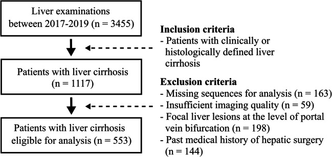
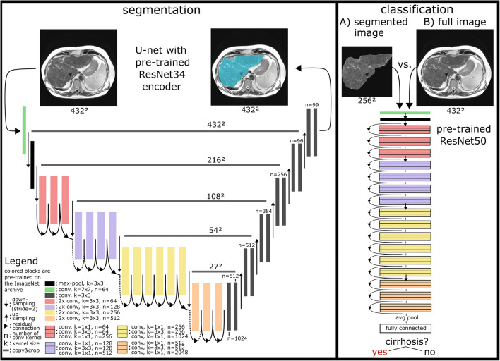
The dataset for this retrospective analysis consisted of 713 (343 female) patients who underwent liver MRI between 2017 and 2019. In total, 553 of these subjects had a confirmed diagnosis of liver cirrhosis, while the remainder had no history of liver disease. T2-weighted MRI slices at the level of the caudate lobe were manually exported for DTL analysis. Data were randomly split into training, validation, and test sets (70%/15%/15%). A ResNet50 convolutional neural network (CNN) pre-trained on the ImageNet archive was used for cirrhosis detection with and without upstream liver segmentation. Classification performance for detection of liver cirrhosis was compared to two radiologists with different levels of experience (4-year resident, board-certified radiologist). Segmentation was performed using a U-Net architecture built on a pre-trained ResNet34 encoder. Differences in classification accuracy were assessed by the χ-test.

RESULTS

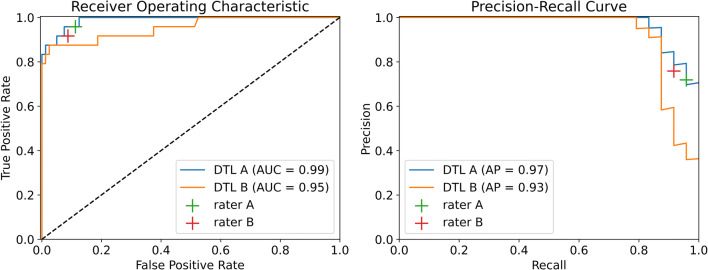
Dice coefficients for automatic segmentation were above 0.98 for both validation and test data. The classification accuracy of liver cirrhosis on validation (vACC) and test (tACC) data for the DTL pipeline with upstream liver segmentation (vACC = 0.99, tACC = 0.96) was significantly higher compared to the resident (vACC = 0.88, p < 0.01; tACC = 0.91, p = 0.01) and to the board-certified radiologist (vACC = 0.96, p < 0.01; tACC = 0.90, p < 0.01).

CONCLUSION

This proof-of-principle study demonstrates the potential of DTL for detecting cirrhosis based on standard T2-weighted MRI. The presented method for image-based diagnosis of liver cirrhosis demonstrated expert-level classification accuracy.

KEY POINTS

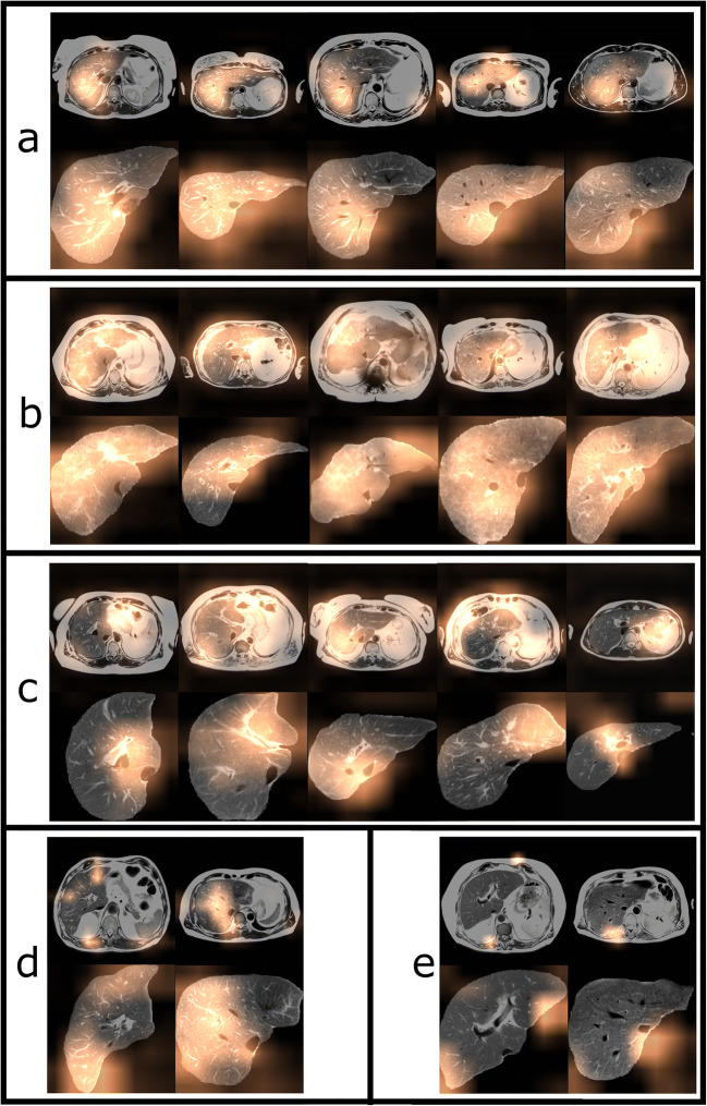
• A pipeline consisting of two convolutional neural networks (CNNs) pre-trained on an extensive natural image database (ImageNet archive) enables detection of liver cirrhosis on standard T2-weighted MRI. • High classification accuracy can be achieved even without altering the pre-trained parameters of the convolutional neural networks. • Other abdominal structures apart from the liver were relevant for detection when the network was trained on unsegmented images.

摘要

目的

探究深度迁移学习(DTL)在从临床 MRI 检测肝硬化中的诊断性能。

方法

本回顾性分析的数据集中包含 713 名(343 名女性)于 2017 年至 2019 年期间进行肝脏 MRI 检查的患者。其中,553 名患者被确诊为肝硬化,其余患者无肝脏疾病史。在尾状叶水平手动导出 T2 加权 MRI 切片进行 DTL 分析。数据随机分为训练集、验证集和测试集(70%/15%/15%)。使用在 ImageNet 存档上预先训练的 ResNet50 卷积神经网络(CNN)进行肝硬化检测,有无上游肝脏分割。将两名具有不同经验水平(4 年住院医师、董事会认证放射科医师)的放射科医生的分类性能与检测肝硬化的性能进行比较。分割使用基于预训练 ResNet34 编码器的 U-Net 架构进行。通过 χ 检验评估分类准确性的差异。

结果

验证(vACC)和测试(tACC)数据中自动分割的 Dice 系数均高于 0.98。具有上游肝脏分割的 DTL 管道(vACC=0.99,tACC=0.96)的肝硬化分类准确性(vACC)在验证(vACC)和测试(tACC)数据中明显高于住院医师(vACC=0.88,p<0.01;tACC=0.91,p=0.01)和董事会认证放射科医师(vACC=0.96,p<0.01;tACC=0.90,p<0.01)。

结论

本研究初步证明了基于标准 T2 加权 MRI 进行 DTL 检测肝硬化的潜力。所提出的基于图像的肝硬化诊断方法达到了专家级别的分类准确性。

重点

• 由两个在大型自然图像数据库(ImageNet 存档)上预先训练的卷积神经网络(CNN)组成的管道可在标准 T2 加权 MRI 上检测肝硬化。• 即使不改变卷积神经网络的预训练参数,也能实现高分类准确性。• 当网络在未分割的图像上进行训练时,除了肝脏之外的其他腹部结构对于检测也很重要。

https://cdn.ncbi.nlm.nih.gov/pmc/blobs/59ed/8523404/c47e25327ef2/330_2021_7858_Fig4_HTML.jpg
https://cdn.ncbi.nlm.nih.gov/pmc/blobs/59ed/8523404/74df3a3847c5/330_2021_7858_Fig1_HTML.jpg
https://cdn.ncbi.nlm.nih.gov/pmc/blobs/59ed/8523404/0caf0ea113b1/330_2021_7858_Fig2_HTML.jpg
https://cdn.ncbi.nlm.nih.gov/pmc/blobs/59ed/8523404/201a46533c21/330_2021_7858_Fig3_HTML.jpg
https://cdn.ncbi.nlm.nih.gov/pmc/blobs/59ed/8523404/c47e25327ef2/330_2021_7858_Fig4_HTML.jpg

相似文献

1
Detection of liver cirrhosis in standard T2-weighted MRI using deep transfer learning.使用深度迁移学习在标准 T2 加权 MRI 中检测肝硬化。
Eur Radiol. 2021 Nov;31(11):8807-8815. doi: 10.1007/s00330-021-07858-1. Epub 2021 May 11.
2
Deep learning supports the differentiation of alcoholic and other-than-alcoholic cirrhosis based on MRI.深度学习支持基于 MRI 区分酒精性和非酒精性肝硬化。
Sci Rep. 2022 May 18;12(1):8297. doi: 10.1038/s41598-022-12410-2.
3
Semiautomated segmentation of hepatocellular carcinoma tumors with MRI using convolutional neural networks.使用卷积神经网络对 MRI 中的肝细胞癌肿瘤进行半自动分割。
Eur Radiol. 2023 Sep;33(9):6020-6032. doi: 10.1007/s00330-023-09613-0. Epub 2023 Apr 18.
4
Semantic segmentation of cerebrospinal fluid and brain volume with a convolutional neural network in pediatric hydrocephalus-transfer learning from existing algorithms.基于卷积神经网络的小儿脑积水脑积水分割及脑容量计算——从现有算法进行迁移学习。
Acta Neurochir (Wien). 2020 Oct;162(10):2463-2474. doi: 10.1007/s00701-020-04447-x. Epub 2020 Jun 25.
5
Ultrasonic Assessment of Liver Fibrosis Using One-Dimensional Convolutional Neural Networks Based on Frequency Spectra of Radiofrequency Signals with Deep Learning Segmentation of Liver Regions in B-Mode Images: A Feasibility Study.基于深度学习的 B 型超声图像肝脏区域分割的射频信号频域一维卷积神经网络对肝纤维化的超声评估:一项可行性研究。
Sensors (Basel). 2024 Aug 26;24(17):5513. doi: 10.3390/s24175513.
6
MABAL: a Novel Deep-Learning Architecture for Machine-Assisted Bone Age Labeling.MABAL:一种用于机器辅助骨龄标注的新型深度学习架构。
J Digit Imaging. 2018 Aug;31(4):513-519. doi: 10.1007/s10278-018-0053-3.
7
Domain- and task-specific transfer learning for medical segmentation tasks.面向医学分割任务的域和任务特定迁移学习。
Comput Methods Programs Biomed. 2022 Feb;214:106539. doi: 10.1016/j.cmpb.2021.106539. Epub 2021 Nov 23.
8
Combined Transfer Learning and Test-Time Augmentation Improves Convolutional Neural Network-Based Semantic Segmentation of Prostate Cancer from Multi-Parametric MR Images.联合迁移学习和测试时增强提高了基于卷积神经网络的多参数磁共振图像前列腺癌的语义分割。
Comput Methods Programs Biomed. 2021 Oct;210:106375. doi: 10.1016/j.cmpb.2021.106375. Epub 2021 Aug 28.
9
Deep morphology aided diagnosis network for segmentation of carotid artery vessel wall and diagnosis of carotid atherosclerosis on black-blood vessel wall MRI.基于深度学习的形态学辅助诊断网络在黑血 MRI 颈动脉血管壁分段及颈动脉粥样硬化诊断中的应用
Med Phys. 2019 Dec;46(12):5544-5561. doi: 10.1002/mp.13739. Epub 2019 Oct 14.
10
Machine Segmentation of Pelvic Anatomy in MRI-Assisted Radiosurgery (MARS) for Prostate Cancer Brachytherapy.MRI 辅助放射外科(MARS)中前列腺癌近距离放射治疗的骨盆解剖结构的机器分割。
Int J Radiat Oncol Biol Phys. 2020 Dec 1;108(5):1292-1303. doi: 10.1016/j.ijrobp.2020.06.076. Epub 2020 Jul 4.

引用本文的文献

1
Advanced liver fibrosis detection using a two-stage deep learning approach on standard T2-weighted MRI.在标准T2加权磁共振成像(MRI)上采用两阶段深度学习方法进行晚期肝纤维化检测。
Abdom Radiol (NY). 2025 Aug 19. doi: 10.1007/s00261-025-05160-y.
2
When liver disease diagnosis encounters deep learning: Analysis, challenges, and prospects.当肝脏疾病诊断遇上深度学习:分析、挑战与展望。
ILIVER. 2023 Mar 4;2(1):73-87. doi: 10.1016/j.iliver.2023.02.002. eCollection 2023 Mar.
3
Enhancing Radiologist Productivity with Artificial Intelligence in Magnetic Resonance Imaging (MRI): A Narrative Review.

本文引用的文献

1
Automatic Hip Fracture Identification and Functional Subclassification with Deep Learning.基于深度学习的髋部骨折自动识别与功能亚分类
Radiol Artif Intell. 2020 Mar 25;2(2):e190023. doi: 10.1148/ryai.2020190023. eCollection 2020 Mar.
2
Automated CT and MRI Liver Segmentation and Biometry Using a Generalized Convolutional Neural Network.使用广义卷积神经网络的肝脏自动CT和MRI分割与生物测量
Radiol Artif Intell. 2019 Mar;1(2). doi: 10.1148/ryai.2019180022. Epub 2019 Mar 27.
3
FastSurfer - A fast and accurate deep learning based neuroimaging pipeline.
利用人工智能提高磁共振成像(MRI)中放射科医生的工作效率:一篇叙述性综述。
Diagnostics (Basel). 2025 Apr 30;15(9):1146. doi: 10.3390/diagnostics15091146.
4
Deep transfer learning radiomics for distinguishing sinonasal malignancies: a preliminary MRI study.用于鉴别鼻窦恶性肿瘤的深度迁移学习放射组学:一项初步的MRI研究
Future Oncol. 2025 Apr;21(8):975-982. doi: 10.1080/14796694.2025.2469486. Epub 2025 Feb 24.
5
MRI-based deep learning radiomics to differentiate dual-phenotype hepatocellular carcinoma from HCC and intrahepatic cholangiocarcinoma: a multicenter study.基于磁共振成像的深度学习影像组学用于鉴别双表型肝细胞癌与肝细胞癌及肝内胆管癌:一项多中心研究
Insights Imaging. 2025 Jan 29;16(1):27. doi: 10.1186/s13244-025-01904-y.
6
AI in Hepatology: Revolutionizing the Diagnosis and Management of Liver Disease.人工智能在肝病学中的应用:革新肝病的诊断与管理
J Clin Med. 2024 Dec 22;13(24):7833. doi: 10.3390/jcm13247833.
7
Fully automated MRI-based convolutional neural network for noninvasive diagnosis of cirrhosis.基于磁共振成像(MRI)的全自动化卷积神经网络用于肝硬化的无创诊断
Insights Imaging. 2024 Dec 12;15(1):298. doi: 10.1186/s13244-024-01872-9.
8
Deep Learning Virtual Contrast-Enhanced T1 Mapping for Contrast-Free Myocardial Extracellular Volume Assessment.深度学习虚拟对比增强 T1 映射在无对比剂心肌细胞外容积评估中的应用。
J Am Heart Assoc. 2024 Oct;13(19):e035599. doi: 10.1161/JAHA.124.035599. Epub 2024 Sep 30.
9
Artificial intelligence techniques in liver cancer.肝癌中的人工智能技术
Front Oncol. 2024 Sep 3;14:1415859. doi: 10.3389/fonc.2024.1415859. eCollection 2024.
10
Diagnostic performance of EfficientNetV2-S method for staging liver fibrosis based on multiparametric MRI.基于多参数磁共振成像的EfficientNetV2-S方法对肝纤维化分期的诊断性能
Heliyon. 2024 Jul 27;10(15):e35115. doi: 10.1016/j.heliyon.2024.e35115. eCollection 2024 Aug 15.
FastSurfer - 一个快速准确的基于深度学习的神经影像学管道。
Neuroimage. 2020 Oct 1;219:117012. doi: 10.1016/j.neuroimage.2020.117012. Epub 2020 Jun 8.
4
On the Interpretability of Artificial Intelligence in Radiology: Challenges and Opportunities.论人工智能在放射学中的可解释性:挑战与机遇
Radiol Artif Intell. 2020 May 27;2(3):e190043. doi: 10.1148/ryai.2020190043.
5
Fully Automated Segmentation of Connective Tissue Compartments for CT-Based Body Composition Analysis: A Deep Learning Approach.基于 CT 的体成分分析的结缔组织分割的全自动方法:深度学习方法。
Invest Radiol. 2020 Jun;55(6):357-366. doi: 10.1097/RLI.0000000000000647.
6
Automated Myocardial T2 and Extracellular Volume Quantification in Cardiac MRI Using Transfer Learning-based Myocardium Segmentation.使用基于迁移学习的心肌分割技术在心脏磁共振成像中实现心肌T2和细胞外容积的自动定量分析。
Radiol Artif Intell. 2020 Jan 29;2(1):e190034. doi: 10.1148/ryai.2019190034.
7
Transfer learning radiomics based on multimodal ultrasound imaging for staging liver fibrosis.基于多模态超声成像的迁移学习放射组学用于分期肝纤维化。
Eur Radiol. 2020 May;30(5):2973-2983. doi: 10.1007/s00330-019-06595-w. Epub 2020 Jan 21.
8
FatSegNet: A fully automated deep learning pipeline for adipose tissue segmentation on abdominal dixon MRI.脂肪分割网络:一种用于腹部狄克逊磁共振成像上脂肪组织分割的全自动深度学习流程。
Magn Reson Med. 2020 Apr;83(4):1471-1483. doi: 10.1002/mrm.28022. Epub 2019 Oct 21.
9
Deep learning with ultrasonography: automated classification of liver fibrosis using a deep convolutional neural network.深度学习与超声:应用深度卷积神经网络对肝纤维化进行自动分类。
Eur Radiol. 2020 Feb;30(2):1264-1273. doi: 10.1007/s00330-019-06407-1. Epub 2019 Sep 2.
10
Radiomics Analysis of Gadoxetic Acid-enhanced MRI for Staging Liver Fibrosis.钆塞酸增强 MRI 影像学组学分析用于肝纤维化分期。
Radiology. 2019 Feb;290(2):380-387. doi: 10.1148/radiol.2018181197. Epub 2018 Dec 4.