van der Voort Sebastian R, Incekara Fatih, Wijnenga Maarten M J, Kapsas Georgios, Gahrmann Renske, Schouten Joost W, Dubbink Hendrikus J, Vincent Arnaud J P E, van den Bent Martin J, French Pim J, Klein Stefan, Smits Marion
Biomedical Imaging Group Rotterdam, Department of Radiology and Nuclear Medicine, Erasmus MC University Medical Centre Rotterdam, Rotterdam, the Netherlands.
Department of Radiology and Nuclear Medicine, Erasmus MC University Medical Centre Rotterdam, Rotterdam, the Netherlands.
Data Brief. 2021 Jun 2;37:107191. doi: 10.1016/j.dib.2021.107191. eCollection 2021 Aug.
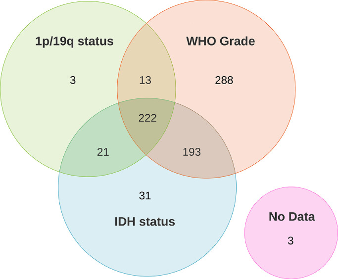
The Erasmus Glioma Database (EGD) contains structural magnetic resonance imaging (MRI) scans, genetic and histological features (specifying the WHO 2016 subtype), and whole tumor segmentations of patients with glioma. Pre-operative MRI data of 774 patients with glioma (281 female, 492 male, 1 unknown, age range 19-86 years) treated at the Erasmus MC between 2008 and 2018 is available. For all patients a pre-contrast T1-weighted, post-contrast T1-weighted, T2-weighted, and T2-weighted FLAIR scan are available, made on a variety of scanners from four different vendors. All scans are registered to a common atlas and defaced. Genetic and histological data consists of the IDH mutation status (available for 467 patients), 1p/19q co-deletion status (available for 259 patients), and grade (available for 716 patients). The full WHO 2016 subtype is available for 415 patients. Manual segmentations are available for 374 patients and automatically generated segmentations are available for 400 patients. The dataset can be used to relate the visual appearance of the tumor on the scan with the genetic and histological features, and to develop automatic segmentation methods.
伊拉斯谟胶质瘤数据库(EGD)包含胶质瘤患者的结构磁共振成像(MRI)扫描、基因和组织学特征(明确WHO 2016亚型)以及全肿瘤分割信息。有2008年至2018年期间在伊拉斯谟医学中心接受治疗的774例胶质瘤患者(281例女性、492例男性、1例性别未知,年龄范围19 - 86岁)的术前MRI数据。所有患者均有平扫T1加权、增强T1加权、T2加权和T2加权液体衰减反转恢复(FLAIR)扫描,这些扫描由来自四个不同供应商的多种扫描仪完成。所有扫描均已配准到一个通用图谱并进行了去标识处理。基因和组织学数据包括异柠檬酸脱氢酶(IDH)突变状态(467例患者可用)、1p/19q共缺失状态(259例患者可用)以及分级(716例患者可用)。415例患者有完整的WHO 2016亚型信息。374例患者有手动分割信息,400例患者有自动生成的分割信息。该数据集可用于将扫描图像上肿瘤的视觉外观与基因和组织学特征相关联,并开发自动分割方法。