Graduate Institute of Oral Biology, School of Dentistry, National Taiwan University, Taipei 10048, Taiwan.
Department of Biomedical Engineering, College of Medicine and College of Engineering, National Taiwan University, Taipei 10617, Taiwan.
Int J Mol Sci. 2021 Jul 20;22(14):7725. doi: 10.3390/ijms22147725.
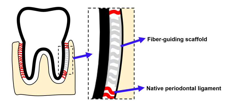
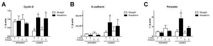
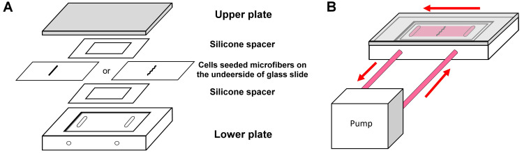
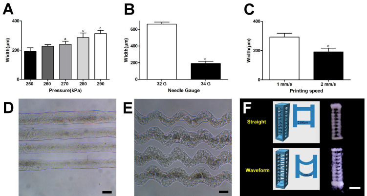
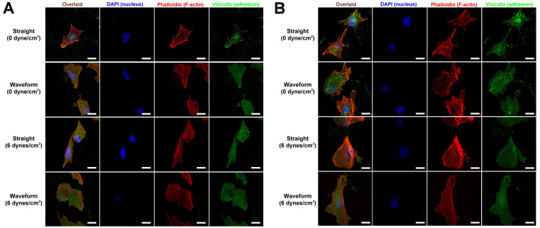
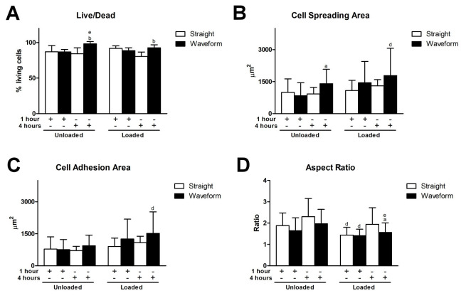
Reconstruction of the periodontal ligament (PDL) to fulfill functional requirement remains a challenge. This study sought to develop a biomimetic microfibrous system capable of withstanding the functional load to assist PDL regeneration. Collagen-based straight and waveform microfibers to guide PDL cell growth were prepared using an extrusion-based bioprinter, and a laminar flow-based bioreactor was used to generate fluidic shear stress. PDL cells were seeded on the respective microfibers with 0 or 6 dynes/cm fluidic shear stress for 1-4 h. The viability, morphology, adhesion pattern, and gene expression levels of PDL cells were assessed. The results revealed that upon bioprinting optimization, collagen-based microfibers were successfully fabricated. The straight microfibers were 189.9 ± 11.44 μm wide and the waveform microfibers were 235.9 ± 11.22 μm wide. Under 6 dynes/cm shear stress, PDL cells were successfully seeded, and cytoskeleton expansion, adhesion, and viability were greater. Cyclin D, E-cadherin, and periostin were upregulated on the waveform microfibers. In conclusion, 3D-printed collagen-based waveform microfibers preserved PDL cell viability and exhibited an enhanced tendency to promote healing and regeneration under shear stress. This approach is promising for the development of a guiding scaffold for PDL regeneration.
重建牙周韧带(PDL)以满足功能需求仍然是一个挑战。本研究旨在开发一种仿生微纤维系统,能够承受功能负荷,以辅助 PDL 再生。使用基于挤出的生物打印机制备基于胶原蛋白的直形和波形微纤维以引导 PDL 细胞生长,并使用层流式生物反应器产生流体剪切力。将 PDL 细胞接种到各自带有 0 或 6 达因/厘米流体剪切力的微纤维上,培养 1-4 小时。评估 PDL 细胞的活力、形态、黏附模式和基因表达水平。结果表明,经过生物打印优化,成功制备了基于胶原蛋白的微纤维。直形微纤维的宽度为 189.9 ± 11.44 μm,波形微纤维的宽度为 235.9 ± 11.22 μm。在 6 达因/厘米剪切力下,成功地接种了 PDL 细胞,细胞骨架扩张、黏附和活力增加。波形微纤维上调了细胞周期蛋白 D、E-钙黏蛋白和骨桥蛋白。总之,3D 打印的基于胶原蛋白的波形微纤维保持了 PDL 细胞的活力,并表现出在剪切力下促进愈合和再生的增强趋势。这种方法有望开发用于 PDL 再生的引导支架。