Müller Dominik, Soto-Rey Iñaki, Kramer Frank
IT-Infrastructure for Translational Medical Research, Faculty of Applied Computer Science, Faculty of Medicine, University of Augsburg, Germany.
Inform Med Unlocked. 2021;25:100681. doi: 10.1016/j.imu.2021.100681. Epub 2021 Jul 27.
The coronavirus disease 2019 (COVID-19) affects billions of lives around the world and has a significant impact on public healthcare. For quantitative assessment and disease monitoring medical imaging like computed tomography offers great potential as alternative to RT-PCR methods. For this reason, automated image segmentation is highly desired as clinical decision support. However, publicly available COVID-19 imaging data is limited which leads to overfitting of traditional approaches.
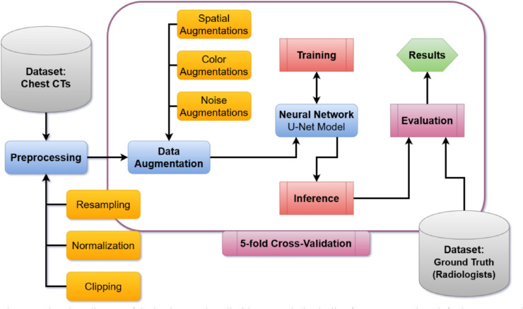
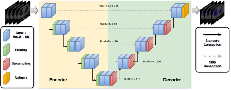
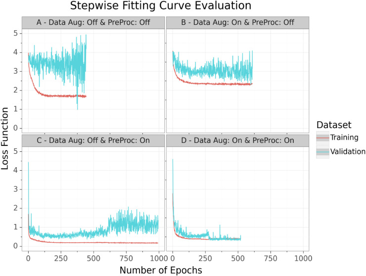
To address this problem, we propose an innovative automated segmentation pipeline for COVID-19 infected regions, which is able to handle small datasets by utilization as variant databases. Our method focuses on on-the-fly generation of unique and random image patches for training by performing several preprocessing methods and exploiting extensive data augmentation. For further reduction of the overfitting risk, we implemented a standard 3D U-Net architecture instead of new or computational complex neural network architectures.
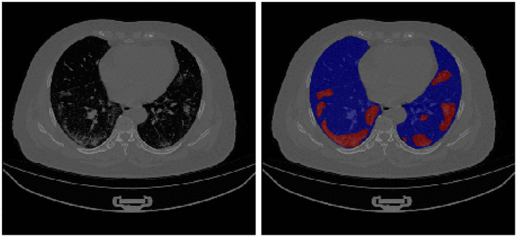
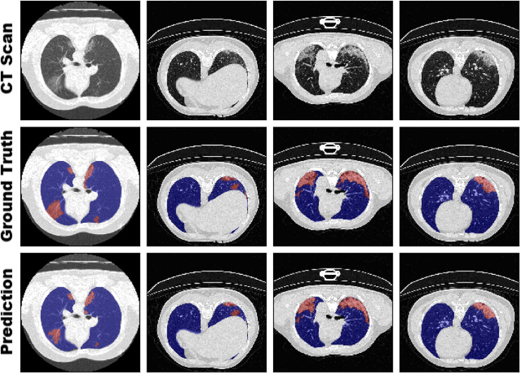
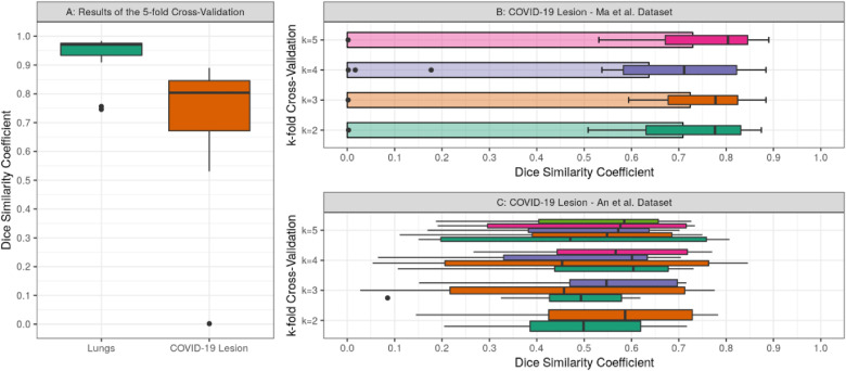
Through a k-fold cross-validation on 20 CT scans as training and validation of COVID-19, we were able to develop a highly accurate as well as robust segmentation model for lungs and COVID-19 infected regions without overfitting on limited data. We performed an in-detail analysis and discussion on the robustness of our pipeline through a sensitivity analysis based on the cross-validation and impact on model generalizability of applied preprocessing techniques. Our method achieved Dice similarity coefficients for COVID-19 infection between predicted and annotated segmentation from radiologists of 0.804 on validation and 0.661 on a separate testing set consisting of 100 patients.
We demonstrated that the proposed method outperforms related approaches, advances the state-of-the-art for COVID-19 segmentation and improves robust medical image analysis based on limited data.
2019冠状病毒病(COVID-19)影响着全球数十亿人的生活,对公共卫生保健产生了重大影响。对于定量评估和疾病监测,计算机断层扫描等医学成像作为逆转录聚合酶链反应(RT-PCR)方法的替代方案具有巨大潜力。因此,作为临床决策支持,自动图像分割非常必要。然而,公开可用的COVID-19成像数据有限,这导致传统方法出现过拟合。
为了解决这个问题,我们提出了一种用于COVID-19感染区域的创新自动分割管道,该管道能够通过用作变体数据库来处理小数据集。我们的方法专注于通过执行几种预处理方法并利用广泛的数据增强来实时生成用于训练的独特随机图像块。为了进一步降低过拟合风险,我们实现了一个标准的3D U-Net架构,而不是新的或计算复杂的神经网络架构。
通过对20例CT扫描进行k折交叉验证作为COVID-19的训练和验证,我们能够开发出一种高度准确且稳健的肺部和COVID-19感染区域分割模型,而不会在有限数据上出现过拟合。我们通过基于交叉验证的敏感性分析以及对应用预处理技术的模型通用性影响,对我们管道的稳健性进行了详细分析和讨论。我们的方法在验证时预测的COVID-19感染与放射科医生标注的分割之间的Dice相似系数为0.804,在由100名患者组成的单独测试集上为0.661。
我们证明了所提出的方法优于相关方法,推动了COVID-19分割的技术水平,并改进了基于有限数据的稳健医学图像分析。