William R. Pritchard Veterinary Medical Teaching Hospital, School of Veterinary Medicine, University of California-Davis, 1 Garrod Drive, Davis, CA, 95695, USA.
Department of Ophthalmology & Vision Science, School of Medicine, U.C. Davis, Sacramento, CA, 95817, USA.
Exp Eye Res. 2021 Nov;212:108754. doi: 10.1016/j.exer.2021.108754. Epub 2021 Sep 11.
To assess age-related changes in the rhesus macaque eye and evaluate them to corresponding human age-related eye disease.
Data from eye exams and imaging tests including intraocular pressure (IOP), lens thickness, axial length, and retinal optical coherence tomography (OCT) images were evaluated from 142 individuals and statistically analyzed for age-related changes. Quantitative autofluorescence (qAF) was measured as was the presence of macular lesions as related to age.
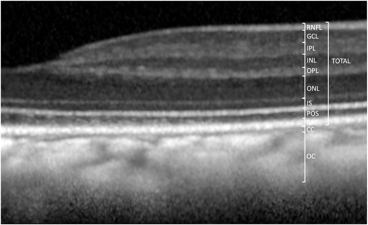
Ages of the 142 rhesus macaques ranged from 0.7 to 29 years (mean = 16.4 years, stdev = 7.5 years). Anterior segment measurements such as IOP, lens thickness, and axial length were acquired. Advanced retinal imaging in the form of optical coherence tomography and qAF were obtained. Quantitative assessments were made and variations by age groups were analyzed to compare with established age-related changes in human eyes. Quantitative analysis of data revealed age-related increase in intraocular pressure (0.165 mm Hg per increase in year of age), ocular biometry (lens thickness 7.2 μm per increase in year of age; and axial length 52.8 μm per increase in year of age), and presence of macular lesions. Age-related changes in thicknesses of retinal layers on OCT were observed and quantified, showing decreased thickness of the retinal ganglion cell layer and inner nuclear layer, and increased thickness of photoreceptor outer segment and choroidal layers. Age was correlated with increased qAF by 1.021 autofluorescence units per increase in year of age.
The rhesus macaque has age-related ocular changes similar to humans. IOP increases with age while retinal ganglion cell layer thickness decreases. Macular lesions develop in some aged animals. Our findings support the concept that rhesus macaques may be useful for the study of important age-related diseases such as glaucoma, macular diseases, and cone disorders, and for development of therapies for these diseases.
评估恒河猴眼睛的年龄相关性变化,并将其与相应的人类年龄相关性眼病进行比较。
从 142 个人的眼部检查和成像测试中评估包括眼压(IOP)、晶状体厚度、眼轴和视网膜光相干断层扫描(OCT)图像的数据,并进行了与年龄相关的变化的统计分析。还测量了定量自发荧光(qAF)以及与年龄相关的黄斑病变的存在情况。
142 只恒河猴的年龄范围为 0.7 至 29 岁(平均 16.4 岁,标准差 7.5 岁)。获得了前节测量值,如 IOP、晶状体厚度和眼轴。获得了高级视网膜成像形式的光相干断层扫描和 qAF。进行了定量评估,并分析了年龄组的变化,以与人类眼睛的既定年龄相关性变化进行比较。数据的定量分析显示,眼压(每年增加 0.165 毫米汞柱)、眼球生物测量(晶状体厚度每年增加 7.2 微米;眼轴每年增加 52.8 微米)和黄斑病变的存在与年龄相关。在 OCT 上观察到并量化了视网膜各层厚度的年龄相关性变化,显示视网膜神经节细胞层和内核层的厚度减少,而光感受器外节和脉络膜层的厚度增加。年龄与 qAF 呈正相关,每年增加 1.021 个自发荧光单位。
恒河猴的眼睛与人类一样存在年龄相关性变化。眼压随年龄增长而增加,而视网膜神经节细胞层厚度减少。一些老年动物出现黄斑病变。我们的发现支持这样一种概念,即恒河猴可能有助于研究重要的年龄相关性疾病,如青光眼、黄斑疾病和锥体细胞疾病,并为这些疾病的治疗开发提供支持。