Laboratory of Neurobiology and Experimental Neurology, Department of Basic Medical Sciences, Faculty of Medicine, La Laguna University, La Laguna, Tenerife, Canary Islands, Spain.
Center for Networked Biomedical Research on Neurodegenerative Diseases (CIBERNED) , Madrid, Spain.
Transl Neurodegener. 2021 Nov 2;10(1):43. doi: 10.1186/s40035-021-00262-1.
The dopaminergic nigrostriatal neurons (DA cells) in healthy people present a slow degeneration with aging, which produces cellular debris throughout life. About 2%-5% of people present rapid cell degeneration of more than 50% of DA cells, which produces Parkinson's disease (PD). Neuroinflammation accelerates the cell degeneration and may be critical for the transition between the slow physiological and the rapid pathological degeneration of DA cells, particularly when it activates microglial cells of the medial forebrain bundle near dopaminergic axons. As synaptic debris produced by DA cell degeneration may trigger the parkinsonian neuroinflammation, this study investigated the removal of axonal debris produced by retrograde degeneration of DA cells, paying particular attention to the relative roles of astrocytes and microglia.
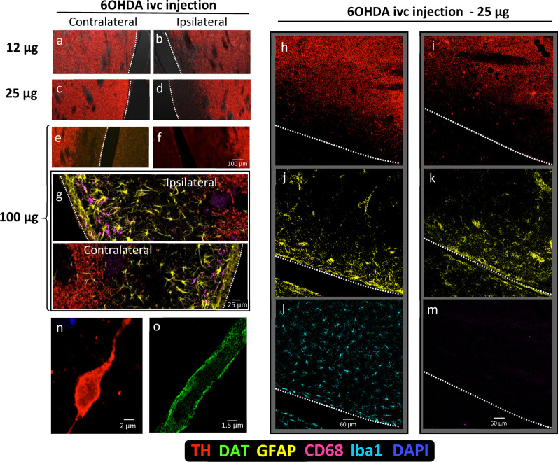
Rats and mice were injected in the lateral ventricles with 6-hydroxydopamine, inducing a degeneration of dopaminergic synapses in the striatum which was not accompanied by non-selective tissue damage, microgliosis or neuroinflammation. The possible retrograde degeneration of dopaminergic axons, and the production and metabolization of DA-cell debris were studied with immunohistochemical methods and analyzed in confocal and electron microscopy images.
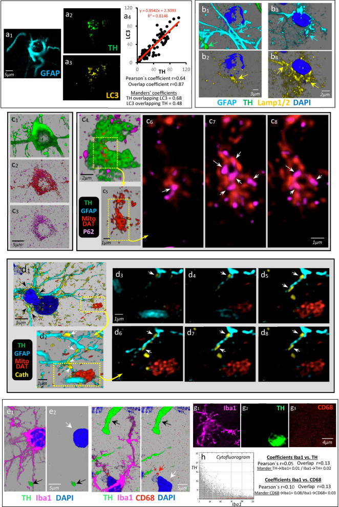
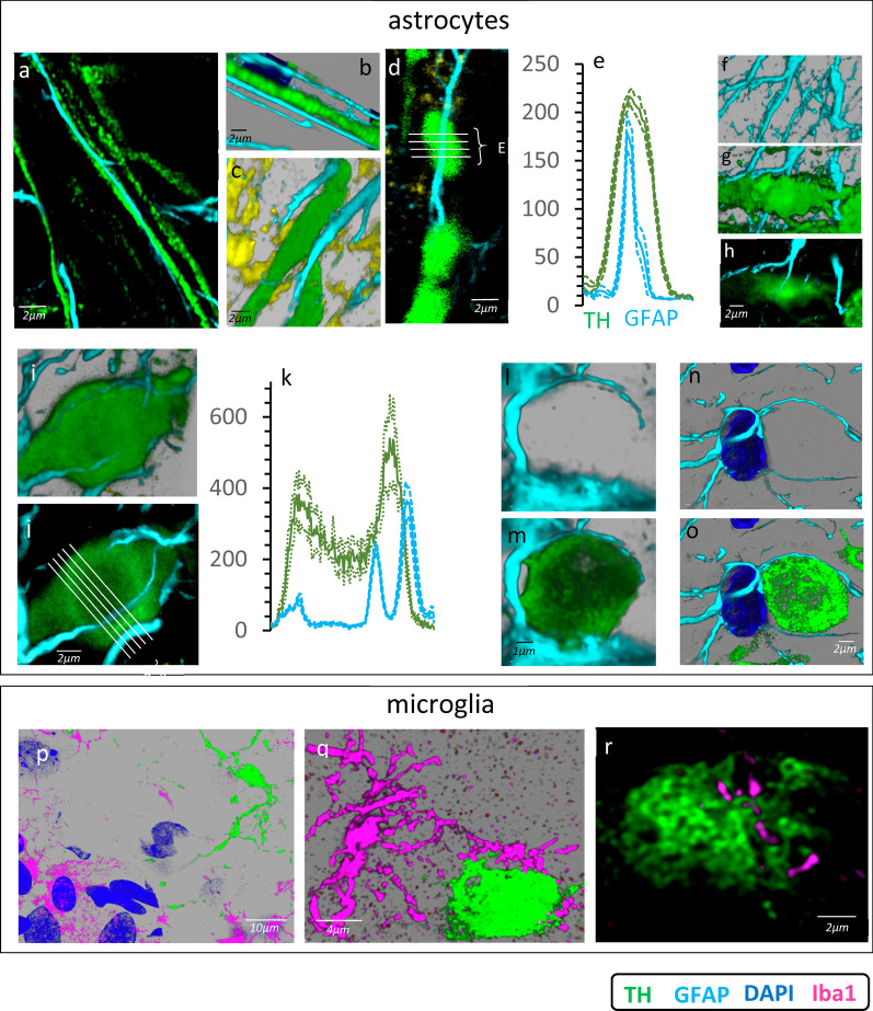
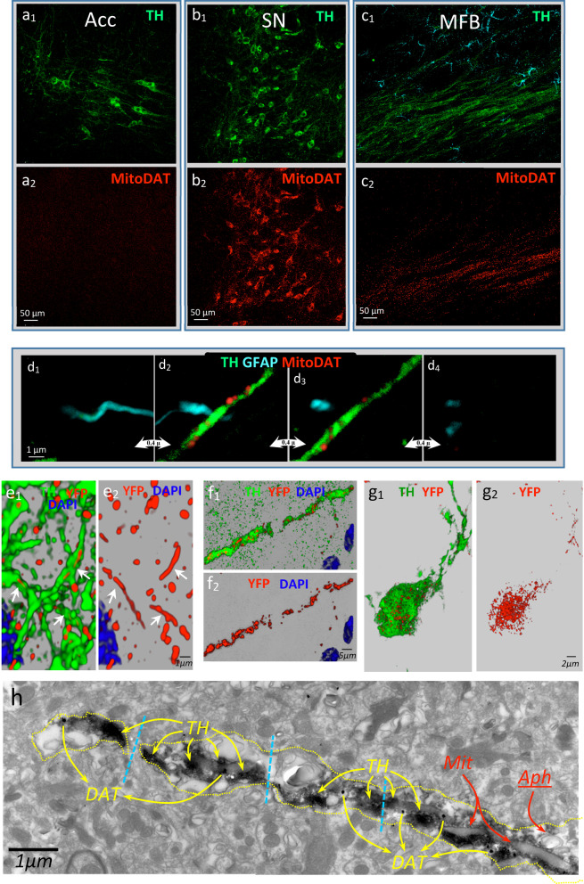
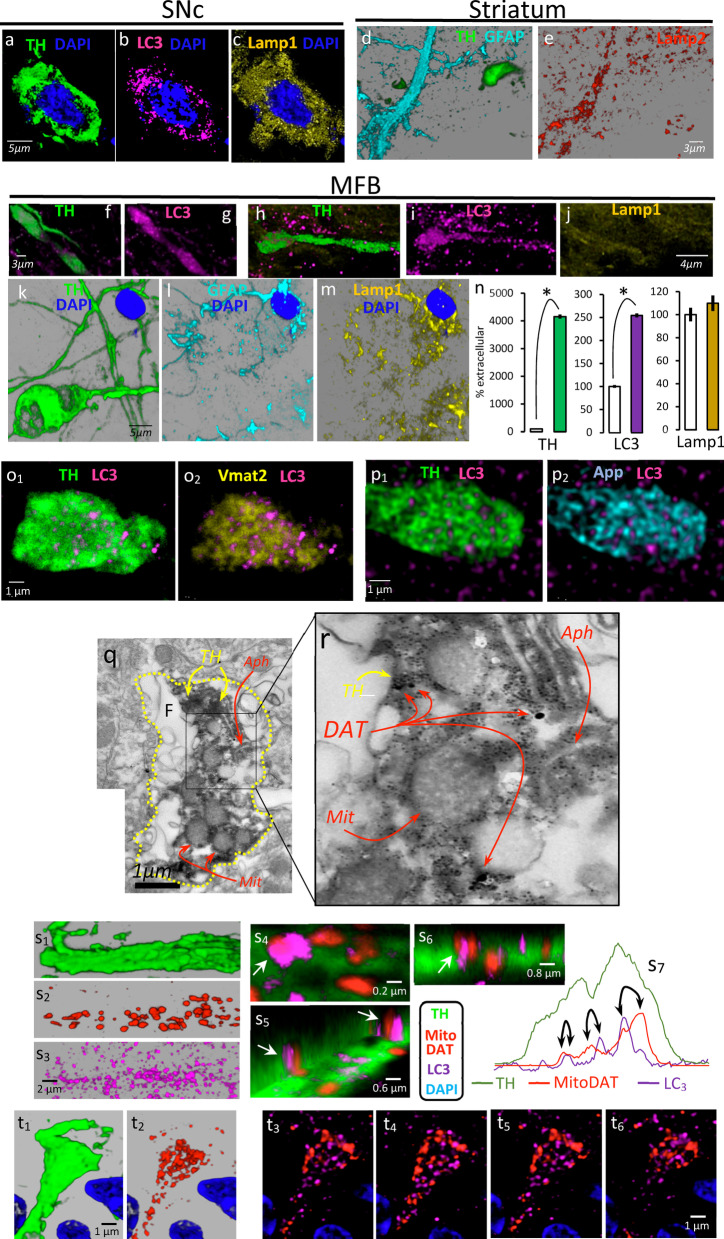
The selective degeneration of dopaminergic synapses in the striatum was followed by a retrograde degeneration of dopaminergic axons whose debris was found within spheroids of the medial forebrain bundle. These spheroids retained mitochondria and most (e.g., tyrosine hydroxylase, the dopamine transporter protein, and amyloid precursor protein) but not all (e.g., α-synuclein) proteins of the degenerating dopaminergic axons. Spheroids showed initial (autophagosomes) but not late (lysosomes) components of autophagy (incomplete autophagy). These spheroids were penetrated by astrocytic processes of the medial forebrain bundle, which provided the lysosomes needed to continue the degradation of dopaminergic debris. Finally, dopaminergic proteins were observed in the cell somata of astrocytes. No microgliosis or microglial phagocytosis of debris was observed in the medial forebrain bundle during the retrograde degeneration of dopaminergic axons.
The present data suggest a physiological role of astrocytic phagocytosis of axonal debris for the medial forebrain bundle astrocytes, which may prevent the activation of microglia and the spread of retrograde axonal degeneration in PD.
健康人群中的多巴胺能黑质纹状体神经元(DA 细胞)随着年龄的增长会出现缓慢退化,从而在一生中产生细胞碎片。大约有 2%-5%的人出现超过 50%的 DA 细胞快速退化,从而导致帕金森病(PD)。神经炎症会加速细胞退化,并且可能对 DA 细胞的缓慢生理退化和快速病理退化之间的转变至关重要,尤其是当它激活靠近多巴胺能轴突的内侧隔束的小胶质细胞时。由于 DA 细胞退化产生的突触碎片可能引发帕金森神经炎症,因此本研究调查了 DA 细胞逆行性退化产生的轴突碎片的清除情况,特别关注星形胶质细胞和小胶质细胞的相对作用。
向大鼠和小鼠的侧脑室注射 6-羟多巴胺,诱导纹状体中多巴胺能突触退化,但不伴有非选择性组织损伤、小胶质细胞增生或神经炎症。用免疫组织化学方法研究多巴胺能轴突的逆行性退化以及 DA 细胞碎片的产生和代谢,并在共聚焦和电子显微镜图像中进行分析。
纹状体中多巴胺能突触的选择性退化后,多巴胺能轴突发生逆行性退化,其碎片在内侧隔束的球体中被发现。这些球体保留了线粒体和大多数(例如,酪氨酸羟化酶、多巴胺转运蛋白和淀粉样前体蛋白)但不是所有(例如,α-突触核蛋白)的退化多巴胺能轴突蛋白。球体显示初始(自噬体)但不是晚期(溶酶体)自噬成分(不完全自噬)。这些球体被内侧隔束的星形胶质细胞突起穿透,这些突起提供了继续降解多巴胺能碎片所需的溶酶体。最后,在星形胶质细胞的细胞体中观察到多巴胺能蛋白。在多巴胺能轴突逆行性退化期间,内侧隔束中未观察到小胶质细胞增生或小胶质细胞吞噬碎片。
目前的数据表明,星形胶质细胞吞噬轴突碎片对于内侧隔束星形胶质细胞具有生理作用,这可能防止小胶质细胞的激活和 PD 中逆行轴突退化的扩散。