Institut Pasteur, Université de Paris, Neural Circuits for Spatial Navigation and Memory, 75015 Paris, France.
Institut Pasteur, Université de Paris, CNRS UMR 3571, Integrative Neurobiology of Cholinergic Systems, 75015 Paris, France; Institut Du Cerveau-Paris Brain Institute-ICM, Sorbonne Université, Inserm U1127, CNRS UMR 7225, 75013 Paris, France.
Cell Rep. 2021 Nov 23;37(8):110035. doi: 10.1016/j.celrep.2021.110035.
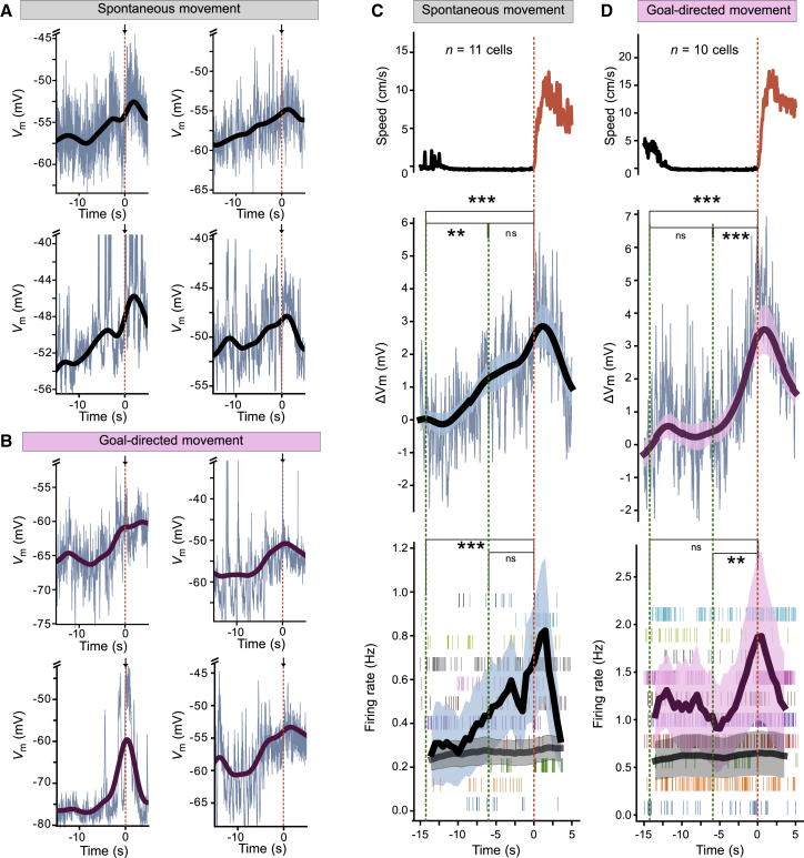
The frontal cortex is essential for organizing voluntary movement. The secondary motor cortex (MOs) is a frontal subregion thought to integrate internal and external inputs before motor action. However, how excitatory and inhibitory synaptic inputs to MOs neurons are integrated preceding movement remains unclear. Here, we address this question by performing in vivo whole-cell recordings from MOs neurons of head-fixed mice moving on a treadmill. We find that principal neurons produce slowly increasing membrane potential and spike ramps preceding spontaneous running. After goal-directed training, ramps show larger amplitudes and accelerated kinetics. Chemogenetic suppression of interneurons combined with modeling suggests that the interplay between parvalbumin-positive (PV+) and somatostatin-positive (SOM+) interneurons, along with principal neuron recurrent connectivity, shape ramping signals. Plasticity of excitatory synapses on SOM+ interneurons can explain the ramp acceleration after training. Altogether, our data reveal that local interneurons differentially control task-dependent ramping signals when MOs neurons integrate inputs preceding movement.
额皮质对于组织自主运动至关重要。次级运动皮质(MOs)是一个额叶亚区,被认为在运动之前整合内部和外部输入。然而,MOs 神经元的兴奋性和抑制性突触输入在运动之前是如何整合的仍不清楚。在这里,我们通过对头固定在跑步机上移动的小鼠的 MOs 神经元进行体内全细胞记录来解决这个问题。我们发现,主要神经元在自发跑动前产生缓慢增加的膜电位和尖峰 ramp。在目标导向训练后,ramp 显示出更大的幅度和更快的动力学。化学遗传抑制中间神经元结合建模表明,PV+和 SOM+中间神经元之间的相互作用,以及主神经元的回传连接,塑造了 ramp 信号。SOM+中间神经元上兴奋性突触的可塑性可以解释训练后 ramp 的加速。总的来说,我们的数据表明,当 MOs 神经元整合运动前的输入时,局部中间神经元以不同的方式控制与任务相关的 ramp 信号。