Cao Yun, Chen Zhaowei, Hu Jijia, Feng Jun, Zhu Zijing, Fan Yanqin, Lin Qiaoxuan, Ding Guohua
Division of Nephrology, Renmin Hospital of Wuhan University, Wuhan, China.
Nephrology and Urology Research Institute of Wuhan University, Wuhan, China.
Front Cell Dev Biol. 2021 Dec 20;9:769213. doi: 10.3389/fcell.2021.769213. eCollection 2021.
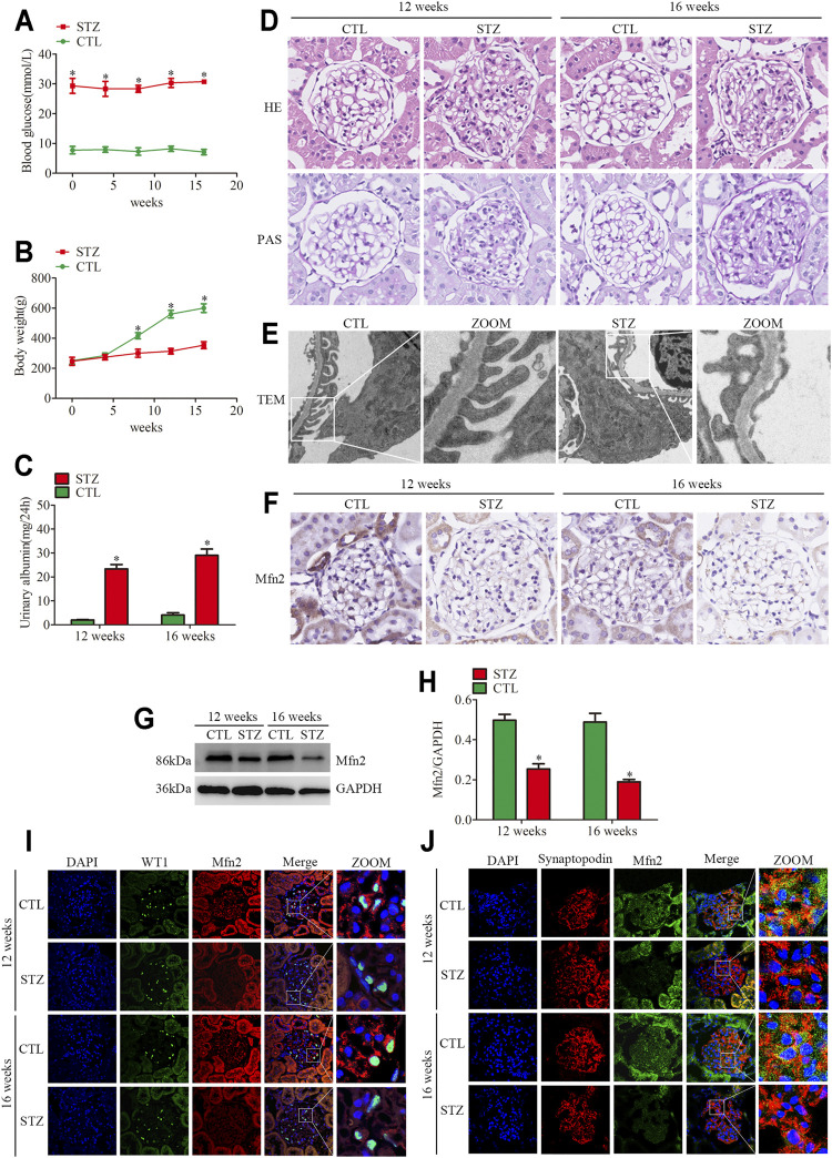
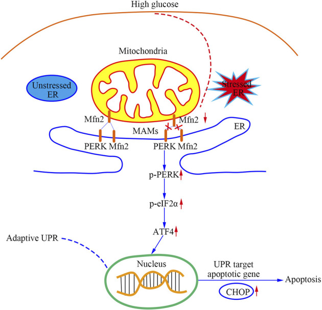
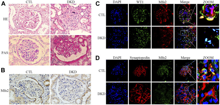
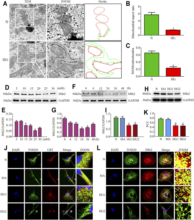
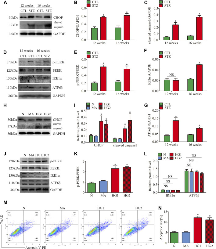
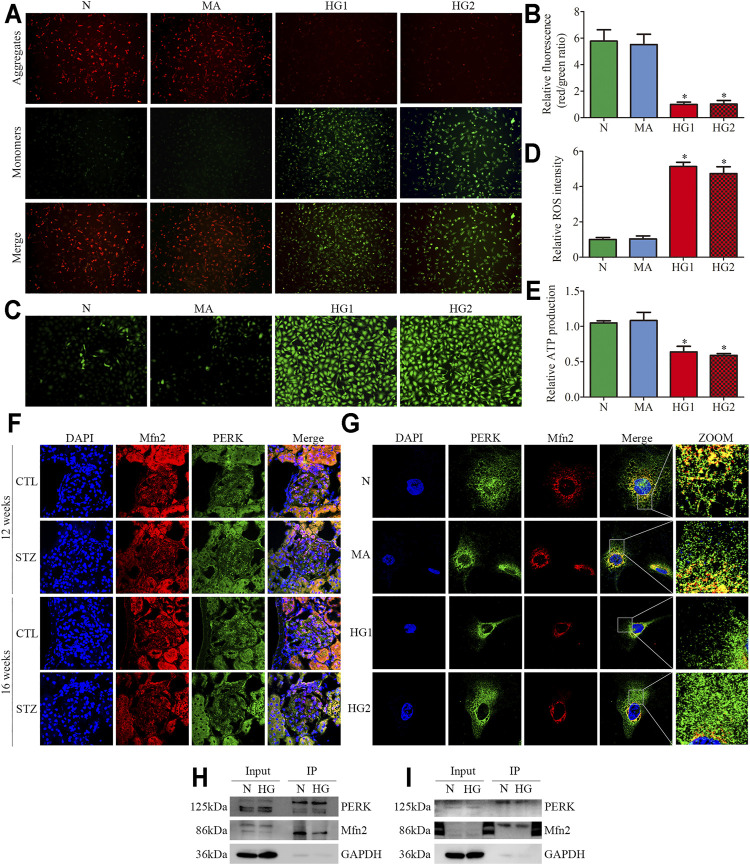
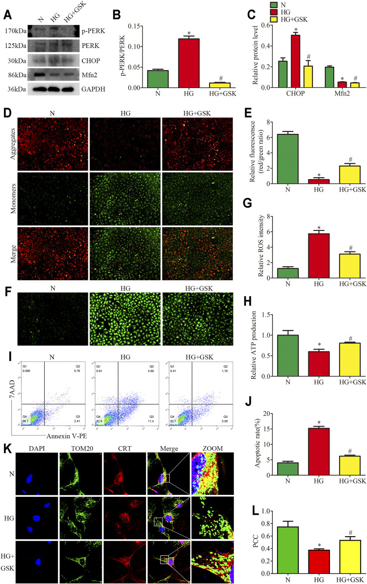
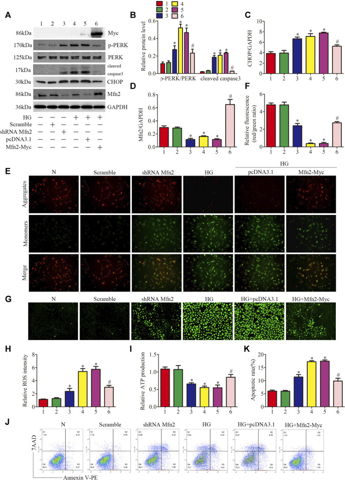
The endoplasmic reticulum (ER) stress and mitochondrial dysfunction in high glucose (HG)-induced podocyte injury have been demonstrated to the progression of diabetic kidney disease (DKD). However, the pathological mechanisms remain equivocal. Mitofusin2 (Mfn2) was initially identified as a dynamin-like protein involved in fusing the outer mitochondrial membrane (OMM). More recently, Mfn2 has been reported to be located at the ER membranes that contact OMM. Mitochondria-associated ER membranes (MAMs) is the intercellular membrane subdomain, which connects the mitochondria and ER through a proteinaceous tether. Here, we observed the suppression of Mfn2 expression in the glomeruli and glomerular podocytes of patients with DKD. Streptozotocin (STZ)-induced diabetic rats exhibited abnormal mitochondrial morphology and MAMs reduction in podocytes, accompanied by decreased expression of Mfn2 and activation of all three unfolded protein response (UPR) pathways (IRE1, ATF6, and PERK). The HG-induced mitochondrial dysfunction, MAMs reduction, and increased apoptosis were accompanied by the downregulation of Mfn2 and activation of the PERK pathway. Mfn2 physically interacts with PERK, and HG promotes a decrease in Mfn2-PERK interaction. In addition, Mfn2-silenced podocytes showed mitochondrial dysfunction, MAMs reduction, activation of PERK pathway, and increased apoptosis. Conversely, all these effects of HG stimulation were alleviated significantly by Mfn2 overexpression. Furthermore, the inhibition of PERK phosphorylation protected mitochondrial functions but did not affect the expression of Mfn2 in HG-treated podocytes. Therefore, this study confirmed that Mfn2 regulates the morphology and functions of MAMs and mitochondria, and exerts anti-apoptotic effects on podocytes by inhibiting the PERK pathway. Hence, the Mfn2-PERK signaling pathway may be a new therapeutic target for preventing podocyte injury in DKD
内质网(ER)应激和高糖(HG)诱导的足细胞损伤中的线粒体功能障碍已被证明与糖尿病肾病(DKD)的进展有关。然而,其病理机制仍不明确。线粒体融合蛋白2(Mfn2)最初被鉴定为一种参与线粒体外膜(OMM)融合的发动蛋白样蛋白。最近,有报道称Mfn2位于与OMM接触的内质网膜上。线粒体相关内质网膜(MAMs)是细胞内膜亚结构域,通过蛋白质连接物连接线粒体和内质网。在这里,我们观察到DKD患者肾小球和肾小球足细胞中Mfn2表达受到抑制。链脲佐菌素(STZ)诱导的糖尿病大鼠足细胞呈现线粒体形态异常和MAMs减少,同时伴有Mfn2表达降低和所有三条未折叠蛋白反应(UPR)途径(IRE1、ATF6和PERK)的激活。HG诱导的线粒体功能障碍、MAMs减少和细胞凋亡增加伴随着Mfn2的下调和PERK途径的激活。Mfn2与PERK发生物理相互作用,HG促进Mfn2-PERK相互作用的减少。此外,Mfn2沉默的足细胞表现出线粒体功能障碍、MAMs减少、PERK途径激活和细胞凋亡增加。相反,Mfn2过表达显著减轻了HG刺激的所有这些影响。此外,抑制PERK磷酸化可保护线粒体功能,但不影响HG处理的足细胞中Mfn2的表达。因此,本研究证实Mfn2调节MAMs和线粒体的形态及功能,并通过抑制PERK途径对足细胞发挥抗凋亡作用。因此,Mfn2-PERK信号通路可能是预防DKD中足细胞损伤的新治疗靶点。