• 文献检索
  • 文档翻译
  • 深度研究
  • 学术资讯
  • Suppr Zotero 插件Zotero 插件
  • 邀请有礼
  • 套餐&价格
  • 历史记录
应用&插件
Suppr Zotero 插件Zotero 插件浏览器插件Mac 客户端Windows 客户端微信小程序
定价
高级版会员购买积分包购买API积分包
服务
文献检索文档翻译深度研究API 文档MCP 服务
关于我们
关于 Suppr公司介绍联系我们用户协议隐私条款
关注我们

Suppr 超能文献

核心技术专利:CN118964589B侵权必究
粤ICP备2023148730 号-1Suppr @ 2026

文献检索

告别复杂PubMed语法,用中文像聊天一样搜索,搜遍4000万医学文献。AI智能推荐,让科研检索更轻松。

立即免费搜索

文件翻译

保留排版,准确专业,支持PDF/Word/PPT等文件格式,支持 12+语言互译。

免费翻译文档

深度研究

AI帮你快速写综述,25分钟生成高质量综述,智能提取关键信息,辅助科研写作。

立即免费体验

一种用于预测伴有腋窝淋巴结转移的乳腺癌腋窝病理完全缓解的临床影像组学模型。

A Clinical-Radiomics Model for Predicting Axillary Pathologic Complete Response in Breast Cancer With Axillary Lymph Node Metastases.

作者信息

Gan Liangyu, Ma Mingming, Liu Yinhua, Liu Qian, Xin Ling, Cheng Yuanjia, Xu Ling, Qin Naishan, Jiang Yuan, Zhang Xiaodong, Wang Xiaoying, Ye Jingming

机构信息

Breast Disease Center, Peking University First Hospital, Beijing, China.

Department of Radiology, Peking University First Hospital, Beijing, China.

出版信息

Front Oncol. 2021 Dec 21;11:786346. doi: 10.3389/fonc.2021.786346. eCollection 2021.

DOI:10.3389/fonc.2021.786346
PMID:34993145
原文链接:https://pmc.ncbi.nlm.nih.gov/articles/PMC8724774/
Abstract

PURPOSE

To develop a clinical-radiomics model based on radiomics features extracted from MRI and clinicopathologic factors for predicting the axillary pathologic complete response (apCR) in breast cancer (BC) patients with axillary lymph node (ALN) metastases.

MATERIALS AND METHODS

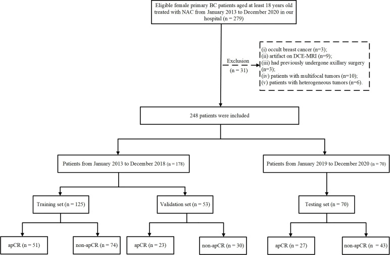
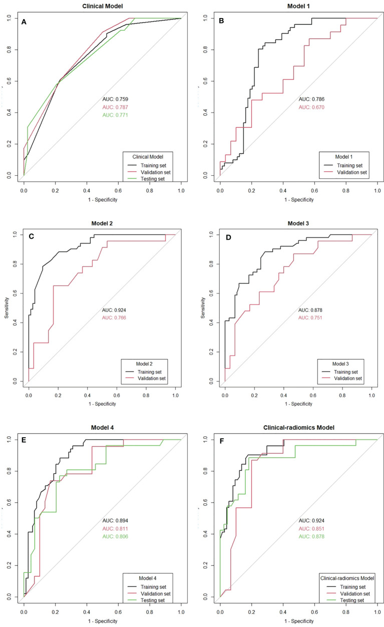
The MR images and clinicopathologic data of 248 eligible invasive BC patients at the Peking University First Hospital from January 2013 to December 2020 were included in this study. All patients received neoadjuvant chemotherapy (NAC), and the presence of ALN metastases was confirmed through cytology pre-NAC. The data from January 2013 to December 2018 were randomly divided into the training and validation sets in a ratio of 7:3, and the data from January 2019 to December 2020 served as the independent testing set. The following three types of prediction models were investigated in this study. 1) A clinical model: the model was built by independently predicting clinicopathologic factors through logistic regression. 2) Radiomics models: we used an automatic segmentation model based on deep learning to segment the axillary areas, visible ALNs, and breast tumors on post-NAC dynamic contrast-enhanced MRI. Radiomics features were then extracted from the region of interest (ROI). Radiomics models were built based on different ROIs or their combination. 3) A clinical-radiomics model: it was built by integrating radiomics signature and independent predictive clinical factors by logistic regression. All models were assessed using a receiver operating characteristic curve analysis and by calculating the area under the curve (AUC).

RESULTS

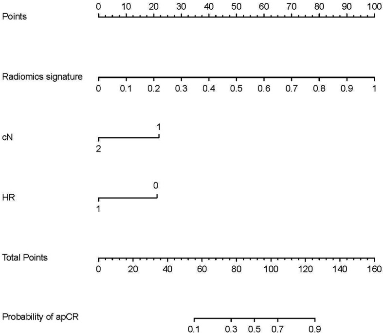
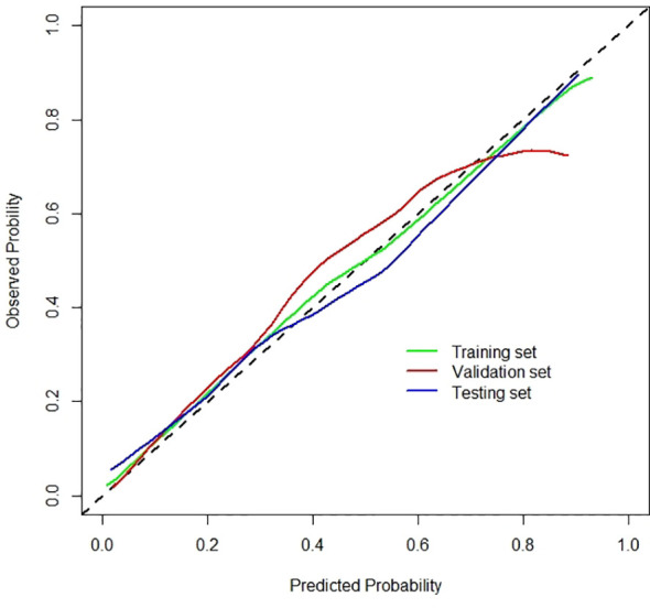
The clinical model yielded AUC values of 0.759, 0.787, and 0.771 in the training, validation, and testing sets, respectively. The radiomics model based on the combination of MRI features of breast tumors and visible ALNs yielded the best AUC values of 0.894, 0.811, and 0.806 in the training, validation, and testing sets, respectively. The clinical-radiomics model yielded AUC values of 0.924, 0.851, and 0.878 in the training, validation, and testing sets, respectively, for predicting apCR.

CONCLUSION

We developed a clinical-radiomics model by integrating radiomics signature and clinical factors to predict apCR in BC patients with ALN metastases post-NAC. It may help the clinicians to screen out apCR patients to avoid lymph node dissection.

摘要

目的

基于从MRI提取的影像组学特征和临床病理因素,开发一种临床影像组学模型,用于预测有腋窝淋巴结(ALN)转移的乳腺癌(BC)患者的腋窝病理完全缓解(apCR)。

材料与方法

本研究纳入了2013年1月至2020年12月在北京大学第一医院的248例符合条件的浸润性BC患者的MR图像和临床病理数据。所有患者均接受了新辅助化疗(NAC),并在NAC前通过细胞学检查确认存在ALN转移。将2013年1月至2018年12月的数据按7:3的比例随机分为训练集和验证集,将2019年1月至2020年12月的数据作为独立测试集。本研究调查了以下三种预测模型。1)临床模型:通过逻辑回归独立预测临床病理因素来构建模型。2)影像组学模型:我们使用基于深度学习的自动分割模型,对NAC后动态对比增强MRI上的腋窝区域、可见的ALN和乳腺肿瘤进行分割。然后从感兴趣区域(ROI)提取影像组学特征。基于不同的ROI或其组合构建影像组学模型。3)临床影像组学模型:通过逻辑回归整合影像组学特征和独立的预测临床因素来构建。所有模型均使用受试者工作特征曲线分析并通过计算曲线下面积(AUC)进行评估。

结果

临床模型在训练集、验证集和测试集中的AUC值分别为0.759、0.787和0.771。基于乳腺肿瘤和可见ALN的MRI特征组合的影像组学模型在训练集、验证集和测试集中分别产生了最佳的AUC值0.894、0.811和0.806。临床影像组学模型在训练集、验证集和测试集中预测apCR的AUC值分别为0.924、0.851和0.878。

结论

我们通过整合影像组学特征和临床因素开发了一种临床影像组学模型,用于预测NAC后有ALN转移的BC患者的apCR。它可能有助于临床医生筛选出apCR患者以避免淋巴结清扫。

https://cdn.ncbi.nlm.nih.gov/pmc/blobs/ec18/8724774/a221ee3d8e55/fonc-11-786346-g007.jpg
https://cdn.ncbi.nlm.nih.gov/pmc/blobs/ec18/8724774/860b56ce29f8/fonc-11-786346-g001.jpg
https://cdn.ncbi.nlm.nih.gov/pmc/blobs/ec18/8724774/2d017b209ed7/fonc-11-786346-g002.jpg
https://cdn.ncbi.nlm.nih.gov/pmc/blobs/ec18/8724774/62177973be84/fonc-11-786346-g003.jpg
https://cdn.ncbi.nlm.nih.gov/pmc/blobs/ec18/8724774/e9f4e0ab8140/fonc-11-786346-g004.jpg
https://cdn.ncbi.nlm.nih.gov/pmc/blobs/ec18/8724774/ccbb53edd810/fonc-11-786346-g005.jpg
https://cdn.ncbi.nlm.nih.gov/pmc/blobs/ec18/8724774/eb863bdf9572/fonc-11-786346-g006.jpg
https://cdn.ncbi.nlm.nih.gov/pmc/blobs/ec18/8724774/a221ee3d8e55/fonc-11-786346-g007.jpg

相似文献

1
A Clinical-Radiomics Model for Predicting Axillary Pathologic Complete Response in Breast Cancer With Axillary Lymph Node Metastases.一种用于预测伴有腋窝淋巴结转移的乳腺癌腋窝病理完全缓解的临床影像组学模型。
Front Oncol. 2021 Dec 21;11:786346. doi: 10.3389/fonc.2021.786346. eCollection 2021.
2
Delta Radiomics Based on MRI for Predicting Axillary Lymph Node Pathologic Complete Response After Neoadjuvant Chemotherapy in Breast Cancer Patients.基于MRI的Delta放射组学预测乳腺癌患者新辅助化疗后腋窝淋巴结病理完全缓解
Acad Radiol. 2025 Jan;32(1):37-49. doi: 10.1016/j.acra.2024.07.052. Epub 2024 Sep 13.
3
Radiomics features based on automatic segmented MRI images: Prognostic biomarkers for triple-negative breast cancer treated with neoadjuvant chemotherapy.基于自动分割 MRI 图像的放射组学特征:新辅助化疗治疗三阴性乳腺癌的预后生物标志物。
Eur J Radiol. 2022 Jan;146:110095. doi: 10.1016/j.ejrad.2021.110095. Epub 2021 Dec 4.
4
Magnetic resonance imaging radiomics predicts preoperative axillary lymph node metastasis to support surgical decisions and is associated with tumor microenvironment in invasive breast cancer: A machine learning, multicenter study.磁共振成像放射组学预测术前腋窝淋巴结转移以支持手术决策,并与浸润性乳腺癌的肿瘤微环境相关:一项机器学习、多中心研究。
EBioMedicine. 2021 Jul;69:103460. doi: 10.1016/j.ebiom.2021.103460. Epub 2021 Jul 4.
5
A Radiomics Model for Preoperative Predicting Sentinel Lymph Node Metastasis in Breast Cancer Based on Dynamic Contrast-Enhanced MRI.基于动态对比增强磁共振成像的乳腺癌前哨淋巴结转移术前预测的放射组学模型
Front Oncol. 2022 Jun 6;12:884599. doi: 10.3389/fonc.2022.884599. eCollection 2022.
6
Development of MRI-Based Deep Learning Signature for Prediction of Axillary Response After NAC in Breast Cancer.基于 MRI 的深度学习特征用于预测乳腺癌新辅助化疗后腋窝反应。
Acad Radiol. 2024 Mar;31(3):800-811. doi: 10.1016/j.acra.2023.10.004. Epub 2023 Oct 31.
7
Prediction of breast cancer and axillary positive-node response to neoadjuvant chemotherapy based on multi-parametric magnetic resonance imaging radiomics models.基于多参数磁共振成像放射组学模型预测乳腺癌和腋窝阳性淋巴结对新辅助化疗的反应。
Breast. 2024 Aug;76:103737. doi: 10.1016/j.breast.2024.103737. Epub 2024 Apr 24.
8
Deep learning radiomics of ultrasonography for comprehensively predicting tumor and axillary lymph node status after neoadjuvant chemotherapy in breast cancer patients: A multicenter study.超声深度学习影像组学用于全面预测乳腺癌患者新辅助化疗后的肿瘤及腋窝淋巴结状态:一项多中心研究
Cancer. 2023 Feb 1;129(3):356-366. doi: 10.1002/cncr.34540. Epub 2022 Nov 19.
9
Longitudinal ultrasound-based AI model predicts axillary lymph node response to neoadjuvant chemotherapy in breast cancer: a multicenter study.基于纵向超声的人工智能模型预测乳腺癌新辅助化疗腋窝淋巴结反应:一项多中心研究。
Eur Radiol. 2024 Nov;34(11):7080-7089. doi: 10.1007/s00330-024-10786-5. Epub 2024 May 10.
10
Prediction of Residual Axillary Nodal Metastasis Following Neoadjuvant Chemotherapy for Breast Cancer: Radiomics Analysis Based on Chest Computed Tomography.基于胸部 CT 的放射组学分析预测乳腺癌新辅助化疗后腋窝残留淋巴结转移
Korean J Radiol. 2023 Jun;24(6):498-511. doi: 10.3348/kjr.2022.0731.

引用本文的文献

1
MRI-based artificial intelligence models for post-neoadjuvant surgery personalization in breast cancer: a narrative review of evidence from Western Pacific.基于MRI的人工智能模型在乳腺癌新辅助治疗后手术个性化中的应用:西太平洋地区证据的叙述性综述
Lancet Reg Health West Pac. 2024 Dec 6;57:101254. doi: 10.1016/j.lanwpc.2024.101254. eCollection 2025 Apr.
2
Research on multi-algorithm and explainable AI techniques for predictive modeling of acute spinal cord injury using multimodal data.使用多模态数据对急性脊髓损伤进行预测建模的多算法与可解释人工智能技术研究。
Sci Rep. 2025 May 29;15(1):18832. doi: 10.1038/s41598-025-93006-4.
3

本文引用的文献

1
Magnetic resonance imaging radiomics predicts preoperative axillary lymph node metastasis to support surgical decisions and is associated with tumor microenvironment in invasive breast cancer: A machine learning, multicenter study.磁共振成像放射组学预测术前腋窝淋巴结转移以支持手术决策,并与浸润性乳腺癌的肿瘤微环境相关:一项机器学习、多中心研究。
EBioMedicine. 2021 Jul;69:103460. doi: 10.1016/j.ebiom.2021.103460. Epub 2021 Jul 4.
2
Construction and Validation of an Immune Infiltration-Related Gene Signature for the Prediction of Prognosis and Therapeutic Response in Breast Cancer.构建和验证一个免疫浸润相关基因特征,用于预测乳腺癌的预后和治疗反应。
Front Immunol. 2021 Apr 27;12:666137. doi: 10.3389/fimmu.2021.666137. eCollection 2021.
3
A combined radiomics and clinical model for preoperative differentiation of intrahepatic cholangiocarcinoma and intrahepatic bile duct stones with cholangitis: a machine learning approach.
一种用于术前鉴别肝内胆管癌与伴胆管炎的肝内胆管结石的联合影像组学和临床模型:一种机器学习方法
Front Oncol. 2025 Mar 17;15:1546940. doi: 10.3389/fonc.2025.1546940. eCollection 2025.
4
Axillary lymph node metastasis in breast cancer: from historical axillary surgery to updated advances in the preoperative diagnosis and axillary management.乳腺癌腋窝淋巴结转移:从传统腋窝手术到术前诊断及腋窝管理的最新进展
BMC Surg. 2025 Feb 27;25(1):81. doi: 10.1186/s12893-025-02802-2.
5
Automatic segmentation-based multi-modal radiomics analysis of US and MRI for predicting disease-free survival of breast cancer: a multicenter study.基于自动分割的多模态放射组学分析在预测乳腺癌无病生存中的应用:一项多中心研究。
Breast Cancer Res. 2024 Nov 12;26(1):157. doi: 10.1186/s13058-024-01909-3.
6
Predicting axillary response to neoadjuvant chemotherapy using peritumoral and intratumoral ultrasound radiomics in breast cancer subtypes.利用瘤周和瘤内超声放射组学预测乳腺癌亚型新辅助化疗的腋窝反应
iScience. 2024 Aug 13;27(9):110716. doi: 10.1016/j.isci.2024.110716. eCollection 2024 Sep 20.
7
Ultrasound and diffuse optical tomography-transformer model for assessing pathological complete response to neoadjuvant chemotherapy in breast cancer.超声与漫射光学断层成像-Transformer 模型评估乳腺癌新辅助化疗的病理完全缓解。
J Biomed Opt. 2024 Jul;29(7):076007. doi: 10.1117/1.JBO.29.7.076007. Epub 2024 Jul 24.
8
A non-invasive artificial intelligence model for identifying axillary pathological complete response to neoadjuvant chemotherapy in breast cancer: a secondary analysis to multicenter clinical trial.一种用于识别乳腺癌新辅助化疗腋窝病理完全缓解的无创人工智能模型:多中心临床试验的二次分析。
Br J Cancer. 2024 Sep;131(4):692-701. doi: 10.1038/s41416-024-02726-3. Epub 2024 Jun 25.
9
Machine learning approaches in the prediction of positive axillary lymph nodes post neoadjuvant chemotherapy using MRI, CT, or ultrasound: A systematic review.使用MRI、CT或超声预测新辅助化疗后腋窝淋巴结阳性的机器学习方法:一项系统综述
Eur J Radiol Open. 2024 Apr 24;12:100561. doi: 10.1016/j.ejro.2024.100561. eCollection 2024 Jun.
10
AI Applications to Breast MRI: Today and Tomorrow.人工智能在乳腺 MRI 中的应用:今天和明天。
J Magn Reson Imaging. 2024 Dec;60(6):2290-2308. doi: 10.1002/jmri.29358. Epub 2024 Apr 5.
Dedicated Axillary MRI-Based Radiomics Analysis for the Prediction of Axillary Lymph Node Metastasis in Breast Cancer.基于专用腋窝MRI的影像组学分析预测乳腺癌腋窝淋巴结转移
Cancers (Basel). 2021 Feb 12;13(4):757. doi: 10.3390/cancers13040757.
4
Development and Validation of a Preoperative Magnetic Resonance Imaging Radiomics-Based Signature to Predict Axillary Lymph Node Metastasis and Disease-Free Survival in Patients With Early-Stage Breast Cancer.基于术前磁共振成像放射组学的signature 模型:预测早期乳腺癌患者腋窝淋巴结转移和无病生存的研究
JAMA Netw Open. 2020 Dec 1;3(12):e2028086. doi: 10.1001/jamanetworkopen.2020.28086.
5
Diagnostic Performance of Noninvasive Imaging for Assessment of Axillary Response After Neoadjuvant Systemic Therapy in Clinically Node-positive Breast Cancer: A Systematic Review and Meta-analysis.临床淋巴结阳性乳腺癌新辅助全身治疗后腋窝反应的无创影像学评估的诊断性能:系统评价和荟萃分析。
Ann Surg. 2021 Apr 1;273(4):694-700. doi: 10.1097/SLA.0000000000004356.
6
Optimizing the Peritumoral Region Size in Radiomics Analysis for Sentinel Lymph Node Status Prediction in Breast Cancer.优化放射组学分析中的肿瘤周围区域大小以预测乳腺癌前哨淋巴结状态。
Acad Radiol. 2022 Jan;29 Suppl 1(Suppl 1):S223-S228. doi: 10.1016/j.acra.2020.10.015. Epub 2020 Nov 5.
7
MRI Radiomics for Assessment of Molecular Subtype, Pathological Complete Response, and Residual Cancer Burden in Breast Cancer Patients Treated With Neoadjuvant Chemotherapy.MRI 放射组学在评估新辅助化疗治疗的乳腺癌患者的分子亚型、病理完全缓解和残留癌负荷中的应用。
Acad Radiol. 2022 Jan;29 Suppl 1(Suppl 1):S145-S154. doi: 10.1016/j.acra.2020.10.020. Epub 2020 Nov 5.
8
A Nomogram Combined Radiomics and Kinetic Curve Pattern as Imaging Biomarker for Detecting Metastatic Axillary Lymph Node in Invasive Breast Cancer.一种结合放射组学和动力学曲线模式的列线图作为检测浸润性乳腺癌腋窝转移淋巴结的影像生物标志物
Front Oncol. 2020 Aug 28;10:1463. doi: 10.3389/fonc.2020.01463. eCollection 2020.
9
Association of higher axillary pathologic complete response rate with breast pathologic complete response after neoadjuvant chemotherapy.新辅助化疗后腋窝病理完全缓解率升高与乳腺病理完全缓解的相关性
Ann Transl Med. 2020 Aug;8(16):992. doi: 10.21037/atm-20-5172.
10
Pharmacokinetic parameters and radiomics model based on dynamic contrast enhanced MRI for the preoperative prediction of sentinel lymph node metastasis in breast cancer.基于动态对比增强 MRI 的药代动力学参数和放射组学模型在乳腺癌前哨淋巴结转移中的术前预测。
Cancer Imaging. 2020 Sep 15;20(1):65. doi: 10.1186/s40644-020-00342-x.