Department of Pathology, Tongji University Affiliated Shanghai Pulmonary Hospital, Shanghai, China.
Shanghai Aitrox Technology Corporation Limited, Shanghai, China.
Mod Pathol. 2022 May;35(5):609-614. doi: 10.1038/s41379-021-00987-4. Epub 2022 Jan 10.
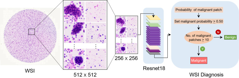
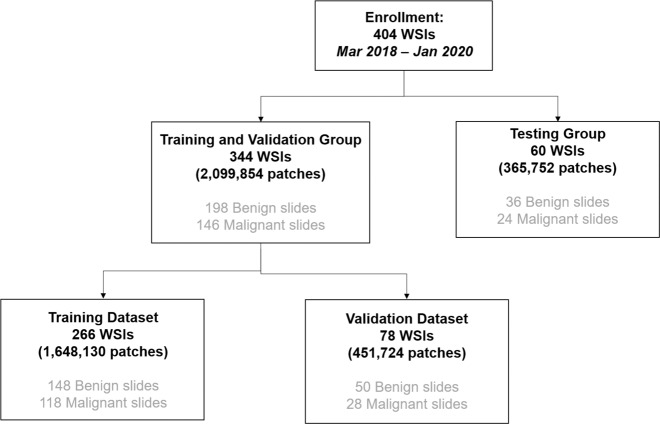
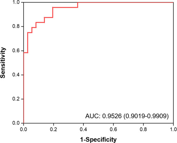
Lung cancer is one of the leading causes of cancer-related death worldwide. Cytology plays an important role in the initial evaluation and diagnosis of patients with lung cancer. However, due to the subjectivity of cytopathologists and the region-dependent diagnostic levels, the low consistency of liquid-based cytological diagnosis results in certain proportions of misdiagnoses and missed diagnoses. In this study, we performed a weakly supervised deep learning method for the classification of benign and malignant cells in lung cytological images through a deep convolutional neural network (DCNN). A total of 404 cases of lung cancer cells in effusion cytology specimens from Shanghai Pulmonary Hospital were investigated, in which 266, 78, and 60 cases were used as the training, validation and test sets, respectively. The proposed method was evaluated on 60 whole-slide images (WSIs) of lung cancer pleural effusion specimens. This study showed that the method had an accuracy, sensitivity, and specificity respectively of 91.67%, 87.50% and 94.44% in classifying malignant and benign lesions (or normal). The area under the receiver operating characteristic (ROC) curve (AUC) was 0.9526 (95% confidence interval (CI): 0.9019-9.9909). In contrast, the average accuracies of senior and junior cytopathologists were 98.34% and 83.34%, respectively. The proposed deep learning method will be useful and may assist pathologists with different levels of experience in the diagnosis of cancer cells on cytological pleural effusion images in the future.
肺癌是全球癌症相关死亡的主要原因之一。细胞学在肺癌患者的初步评估和诊断中发挥着重要作用。然而,由于细胞病理学家的主观性和诊断水平的地区依赖性,液基细胞学诊断结果的一致性较低,导致一定比例的误诊和漏诊。在这项研究中,我们通过深度卷积神经网络(DCNN)对肺癌细胞学图像中的良性和恶性细胞进行了弱监督深度学习方法的分类。共研究了上海肺科医院胸水细胞学标本中的 404 例肺癌细胞,其中 266、78 和 60 例分别作为训练、验证和测试集。该方法在 60 张肺癌胸腔积液标本的全切片图像(WSI)上进行了评估。该研究表明,该方法在区分良恶性病变(或正常)时的准确率、敏感度和特异度分别为 91.67%、87.50%和 94.44%。受试者工作特征(ROC)曲线下面积(AUC)为 0.9526(95%置信区间(CI):0.9019-9.9909)。相比之下,高级和初级细胞病理学家的平均准确率分别为 98.34%和 83.34%。该深度学习方法将是有用的,并可能有助于不同经验水平的病理学家在未来对细胞学胸腔积液图像中的癌细胞进行诊断。