Rao Divya, K Prakashini, Singh Rohit, J Vijayananda
Department of Information and Communication Technology, Manipal Institute of Technology, Manipal Academy of Higher Education, 576104 Manipal, India.
Department of Otorhinolaryngology, Kasturba Medical College, Manipal Academy of Higher Education, 576104 Manipal, India.
Biomed Eng Lett. 2022 Mar 18;12(2):175-183. doi: 10.1007/s13534-022-00221-3. eCollection 2022 May.
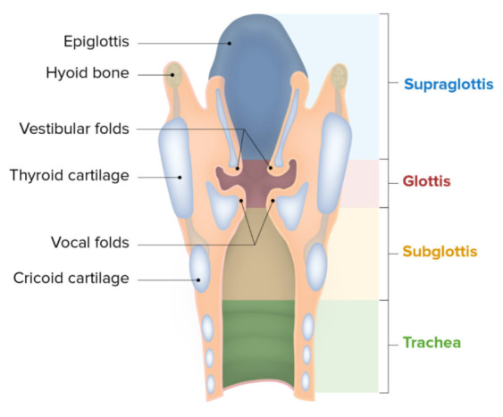
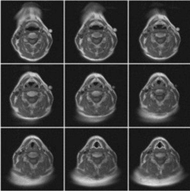
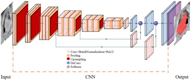
The larynx, or the voice-box, is a common site of occurrence of Head and Neck cancers. Yet, automated segmentation of the larynx has been receiving very little attention. Segmentation of organs is an essential step in cancer treatment-planning. Computed Tomography scans are routinely used to assess the extent of tumor spread in the Head and Neck as they are fast to acquire and tolerant to some movement. This paper reviews various automated detection and segmentation methods used for the larynx on Computed Tomography images. Image registration and deep learning approaches to segmenting the laryngeal anatomy are compared, highlighting their strengths and shortcomings. A list of available annotated laryngeal computed tomography datasets is compiled for encouraging further research. Commercial software currently available for larynx contouring are briefed in our work. We conclude that the lack of standardisation on larynx boundaries and the complexity of the relatively small structure makes automated segmentation of the larynx on computed tomography images a challenge. Reliable computer aided intervention in the contouring and segmentation process will help clinicians easily verify their findings and look for oversight in diagnosis. This review is useful for research that works with artificial intelligence in Head and Neck cancer, specifically that deals with the segmentation of laryngeal anatomy.
The online version contains supplementary material available at 10.1007/s13534-022-00221-3.
喉,即声门,是头颈癌的常见发病部位。然而,喉的自动分割一直很少受到关注。器官分割是癌症治疗计划中的关键步骤。计算机断层扫描(CT)由于采集速度快且能容忍一定程度的运动,常被用于评估头颈肿瘤的扩散范围。本文综述了在CT图像上用于喉的各种自动检测和分割方法。比较了用于分割喉解剖结构的图像配准和深度学习方法,突出了它们的优缺点。编制了一份可用的带注释的喉CT数据集列表,以鼓励进一步的研究。我们的工作还简要介绍了目前可用于喉轮廓勾画的商业软件。我们得出结论,喉边界缺乏标准化以及相对较小结构的复杂性使得在CT图像上对喉进行自动分割成为一项挑战。在轮廓勾画和分割过程中可靠的计算机辅助干预将有助于临床医生轻松验证其发现并查找诊断中的疏漏。这篇综述对于在头颈癌中使用人工智能的研究,特别是涉及喉解剖结构分割的研究很有用。
在线版本包含可在10.1007/s13534-022-00221-3获取的补充材料。