College of Information Science and Engineering, Xinjiang University, Northwest Road, Shayibake District, Urumqi, 830046, Xinjiang, China.
Key Laboratory of Signal Detection and Processing, Xinjiang University, Urumqi, 830046, China.
BMC Med Inform Decis Mak. 2022 Jul 4;22(1):176. doi: 10.1186/s12911-022-01919-1.
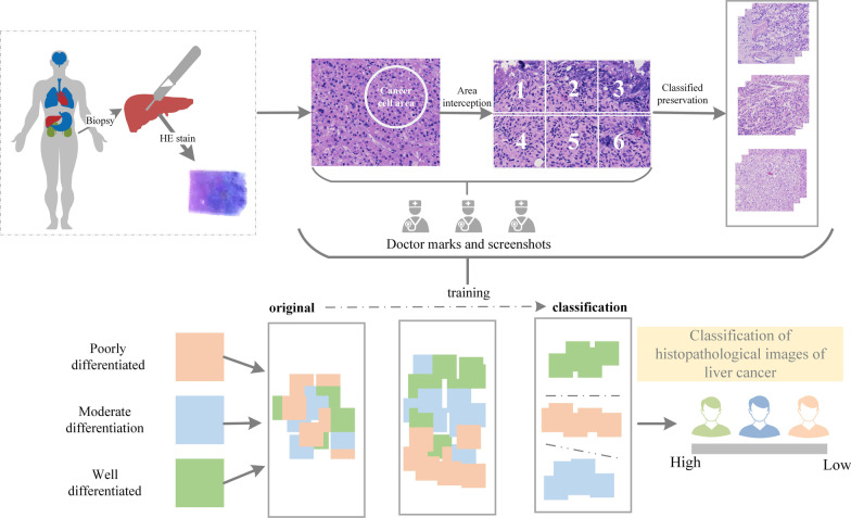
Liver cancer is one of the most common malignant tumors in the world, ranking fifth in malignant tumors. The degree of differentiation can reflect the degree of malignancy. The degree of malignancy of liver cancer can be divided into three types: poorly differentiated, moderately differentiated, and well differentiated. Diagnosis and treatment of different levels of differentiation are crucial to the survival rate and survival time of patients. As the gold standard for liver cancer diagnosis, histopathological images can accurately distinguish liver cancers of different levels of differentiation. Therefore, the study of intelligent classification of histopathological images is of great significance to patients with liver cancer. At present, the classification of histopathological images of liver cancer with different degrees of differentiation has disadvantages such as time-consuming, labor-intensive, and large manual investment. In this context, the importance of intelligent classification of histopathological images is obvious.
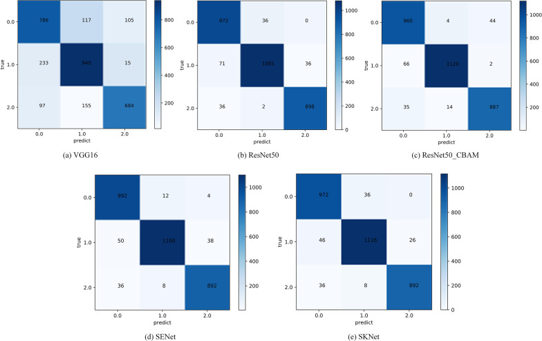
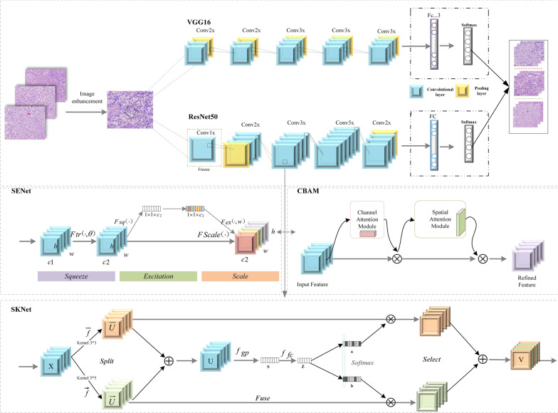
Based on the development of a complete data acquisition scheme, this paper applies the SENet deep learning model to the intelligent classification of all types of differentiated liver cancer histopathological images for the first time, and compares it with the four deep learning models of VGG16, ResNet50, ResNet_CBAM, and SKNet. The evaluation indexes adopted in this paper include confusion matrix, Precision, recall, F1 Score, etc. These evaluation indexes can be used to evaluate the model in a very comprehensive and accurate way.
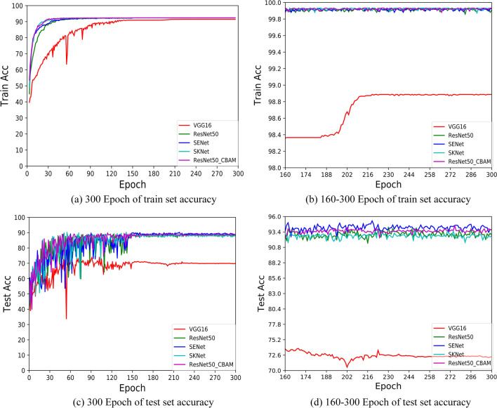
Five different deep learning classification models are applied to collect the data set and evaluate model. The experimental results show that the SENet model has achieved the best classification effect with an accuracy of 95.27%. The model also has good reliability and generalization ability. The experiment proves that the SENet deep learning model has a good application prospect in the intelligent classification of histopathological images.
This study also proves that deep learning has great application value in solving the time-consuming and laborious problems existing in traditional manual film reading, and it has certain practical significance for the intelligent classification research of other cancer histopathological images.
肝癌是世界上最常见的恶性肿瘤之一,在恶性肿瘤中排名第五。分化程度可以反映恶性程度。肝癌的恶性程度可以分为低分化、中分化和高分化三种类型。不同分化程度的诊断和治疗对患者的生存率和生存时间至关重要。作为肝癌诊断的金标准,组织病理学图像可以准确区分不同分化程度的肝癌。因此,研究组织病理学图像的智能分类对肝癌患者具有重要意义。目前,不同分化程度的肝癌组织病理学图像分类存在耗时、费力、人工投入大等缺点。在这种情况下,智能分类组织病理学图像的重要性不言而喻。
本研究基于完整的数据采集方案,首次将 SENet 深度学习模型应用于所有类型分化的肝癌组织病理学图像的智能分类,并与 VGG16、ResNet50、ResNet_CBAM 和 SKNet 四种深度学习模型进行比较。本文采用混淆矩阵、准确率、召回率、F1 分数等评价指标,这些评价指标可以非常全面和准确地评价模型。
将五种不同的深度学习分类模型应用于数据集中进行模型评估。实验结果表明,SENet 模型的分类效果最佳,准确率为 95.27%。该模型还具有良好的可靠性和泛化能力。实验证明,SENet 深度学习模型在组织病理学图像的智能分类中具有良好的应用前景。
本研究还证明,深度学习在解决传统手动读片耗时费力的问题方面具有很大的应用价值,对其他癌症组织病理学图像的智能分类研究具有一定的实际意义。