• 文献检索
  • 文档翻译
  • 深度研究
  • 学术资讯
  • Suppr Zotero 插件Zotero 插件
  • 邀请有礼
  • 套餐&价格
  • 历史记录
应用&插件
Suppr Zotero 插件Zotero 插件浏览器插件Mac 客户端Windows 客户端微信小程序
定价
高级版会员购买积分包购买API积分包
服务
文献检索文档翻译深度研究API 文档MCP 服务
关于我们
关于 Suppr公司介绍联系我们用户协议隐私条款
关注我们

Suppr 超能文献

核心技术专利:CN118964589B侵权必究
粤ICP备2023148730 号-1Suppr @ 2026

文献检索

告别复杂PubMed语法,用中文像聊天一样搜索,搜遍4000万医学文献。AI智能推荐,让科研检索更轻松。

立即免费搜索

文件翻译

保留排版,准确专业,支持PDF/Word/PPT等文件格式,支持 12+语言互译。

免费翻译文档

深度研究

AI帮你快速写综述,25分钟生成高质量综述,智能提取关键信息,辅助科研写作。

立即免费体验

使用 ASHLAR 对组织和肿瘤的高多重化全幻灯片图像进行拼接和配准。

Stitching and registering highly multiplexed whole-slide images of tissues and tumors using ASHLAR.

机构信息

Human Tumor Atlas Network, Harvard Medical School, Boston, MA 02115, USA.

Harvard Ludwig Cancer Center and Laboratory of Systems Pharmacology, Harvard Medical School, Boston, MA 02115, USA.

出版信息

Bioinformatics. 2022 Sep 30;38(19):4613-4621. doi: 10.1093/bioinformatics/btac544.

DOI:10.1093/bioinformatics/btac544
PMID:35972352
原文链接:https://pmc.ncbi.nlm.nih.gov/articles/PMC9525007/
Abstract

MOTIVATION

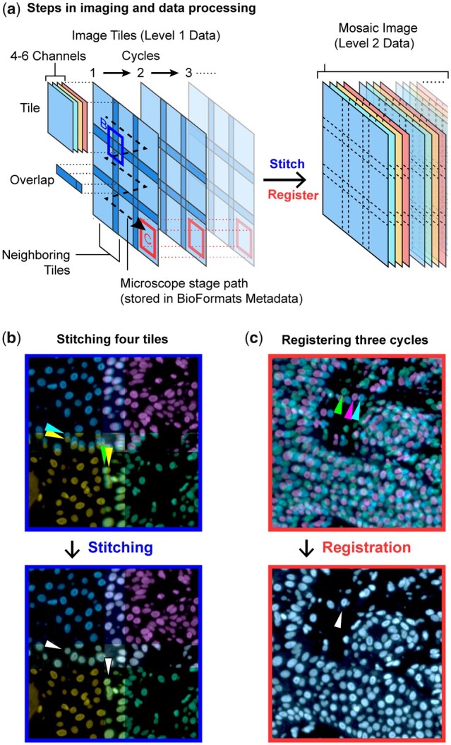
Stitching microscope images into a mosaic is an essential step in the analysis and visualization of large biological specimens, particularly human and animal tissues. Recent approaches to highly multiplexed imaging generate high-plex data from sequential rounds of lower-plex imaging. These multiplexed imaging methods promise to yield precise molecular single-cell data and information on cellular neighborhoods and tissue architecture. However, attaining mosaic images with single-cell accuracy requires robust image stitching and image registration capabilities that are not met by existing methods.

RESULTS

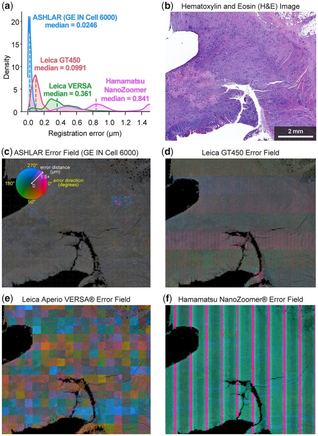
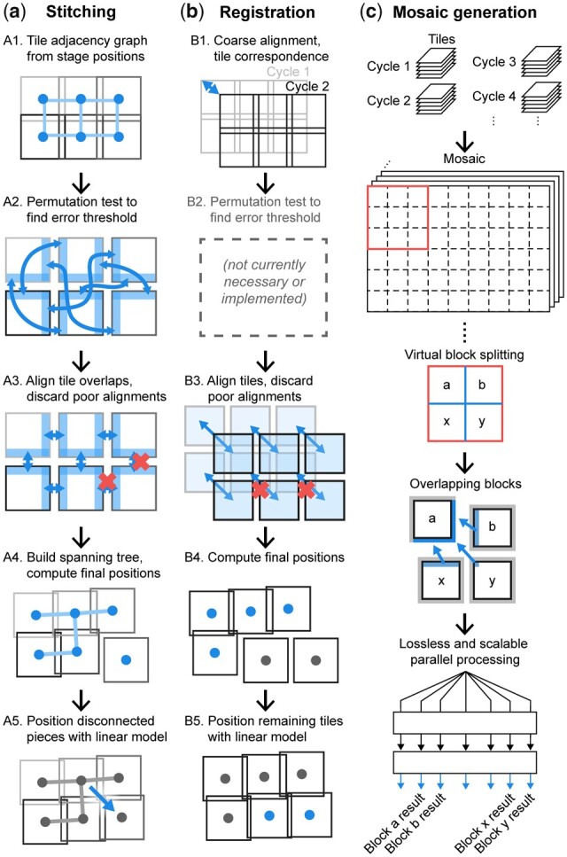
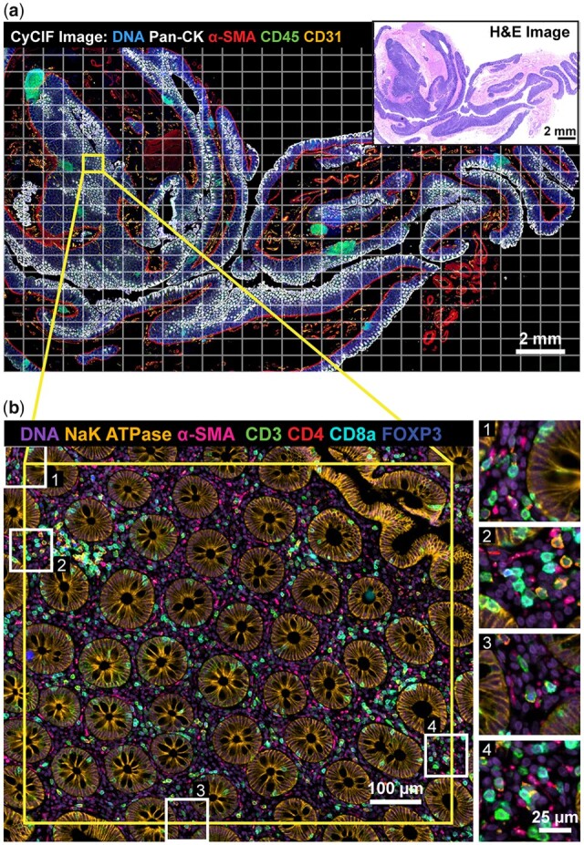
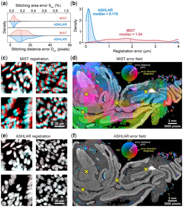
We describe the development and testing of ASHLAR, a Python tool for coordinated stitching and registration of 103 or more individual multiplexed images to generate accurate whole-slide mosaics. ASHLAR reads image formats from most commercial microscopes and slide scanners, and we show that it performs better than existing open-source and commercial software. ASHLAR outputs standard OME-TIFF images that are ready for analysis by other open-source tools and recently developed image analysis pipelines.

AVAILABILITY AND IMPLEMENTATION

ASHLAR is written in Python and is available under the MIT license at https://github.com/labsyspharm/ashlar. The newly published data underlying this article are available in Sage Synapse at https://dx.doi.org/10.7303/syn25826362; the availability of other previously published data re-analyzed in this article is described in Supplementary Table S4. An informational website with user guides and test data is available at https://labsyspharm.github.io/ashlar/.

SUPPLEMENTARY INFORMATION

Supplementary data are available at Bioinformatics online.

摘要

动机

将显微镜图像拼接成马赛克是分析和可视化大型生物样本(尤其是人类和动物组织)的重要步骤。最近的高通量成像方法通过连续多轮低通量成像生成高通量数据。这些高通量成像方法有望提供精确的分子单细胞数据以及关于细胞邻居和组织架构的信息。然而,要实现具有单细胞精度的马赛克图像,需要稳健的图像拼接和图像配准功能,而现有的方法无法满足这些需求。

结果

我们描述了 ASHLAR 的开发和测试,这是一种用于协调拼接和注册 103 个或更多个个体的多路复用图像以生成准确的全幻灯片马赛克的 Python 工具。ASHLAR 可以读取来自大多数商业显微镜和幻灯片扫描仪的图像格式,我们表明它的性能优于现有的开源和商业软件。ASHLAR 输出标准的 OME-TIFF 图像,可用于其他开源工具和最近开发的图像分析管道进行分析。

可用性和实现

ASHLAR 是用 Python 编写的,可在 MIT 许可证下从 https://github.com/labsyspharm/ashlar 获得。本文所依据的新发布的数据可在 Sage Synapse 上 https://dx.doi.org/10.7303/syn25826362 获得;本文重新分析的其他先前发布的数据的可用性在补充表 S4 中描述。一个带有用户指南和测试数据的信息网站可在 https://labsyspharm.github.io/ashlar/ 获得。

补充信息

补充数据可在 Bioinformatics 在线获得。

https://cdn.ncbi.nlm.nih.gov/pmc/blobs/bd82/9525007/982d5e60cdee/btac544f6.jpg
https://cdn.ncbi.nlm.nih.gov/pmc/blobs/bd82/9525007/e2f43642f4a0/btac544f1.jpg
https://cdn.ncbi.nlm.nih.gov/pmc/blobs/bd82/9525007/4fc72bc722df/btac544f2.jpg
https://cdn.ncbi.nlm.nih.gov/pmc/blobs/bd82/9525007/7589ccd1c69f/btac544f3.jpg
https://cdn.ncbi.nlm.nih.gov/pmc/blobs/bd82/9525007/b7ba6a37fa22/btac544f4.jpg
https://cdn.ncbi.nlm.nih.gov/pmc/blobs/bd82/9525007/64eaf2855096/btac544f5.jpg
https://cdn.ncbi.nlm.nih.gov/pmc/blobs/bd82/9525007/982d5e60cdee/btac544f6.jpg

相似文献

1
Stitching and registering highly multiplexed whole-slide images of tissues and tumors using ASHLAR.使用 ASHLAR 对组织和肿瘤的高多重化全幻灯片图像进行拼接和配准。
Bioinformatics. 2022 Sep 30;38(19):4613-4621. doi: 10.1093/bioinformatics/btac544.
2
Automated image mosaics by non-automated light microscopes: the MicroMos software tool.非自动化光学显微镜的自动图像拼接:MicroMos 软件工具。
J Microsc. 2013 Dec;252(3):226-50. doi: 10.1111/jmi.12084. Epub 2013 Sep 20.
3
Correction of image distortion in large-field ssEM stitching by an unsupervised intermediate-space solving network.通过无监督中间空间求解网络校正大视场 ssEM 拼接中的图像变形。
Bioinformatics. 2022 Oct 14;38(20):4797-4805. doi: 10.1093/bioinformatics/btac566.
4
Quantifying and correcting slide-to-slide variation in multiplexed immunofluorescence images.定量和校正多重免疫荧光图像的幻灯片到幻灯片的变化。
Bioinformatics. 2022 Mar 4;38(6):1700-1707. doi: 10.1093/bioinformatics/btab877.
5
CellAnimation: an open source MATLAB framework for microscopy assays.细胞动画:一个用于显微镜检测的开源 MATLAB 框架。
Bioinformatics. 2012 Jan 1;28(1):138-9. doi: 10.1093/bioinformatics/btr633. Epub 2011 Nov 24.
6
Virtual alignment of pathology image series for multi-gigapixel whole slide images.多吉帕像素全玻片图像的病理学图像系列的虚拟配准。
Nat Commun. 2023 Jul 26;14(1):4502. doi: 10.1038/s41467-023-40218-9.
7
PreMosa: extracting 2D surfaces from 3D microscopy mosaics.PreMosa:从 3D 显微镜镶嵌图像中提取 2D 表面。
Bioinformatics. 2017 Aug 15;33(16):2563-2569. doi: 10.1093/bioinformatics/btx195.
8
NRStitcher: non-rigid stitching of terapixel-scale volumetric images.NRStitcher:万亿像素级容积图像的非刚性拼接。
Bioinformatics. 2019 Dec 15;35(24):5290-5297. doi: 10.1093/bioinformatics/btz423.
9
A SIMPLI (Single-cell Identification from MultiPLexed Images) approach for spatially-resolved tissue phenotyping at single-cell resolution.一种基于 SIMPLI(从多重成像中鉴定单细胞)的方法,可实现单细胞分辨率的空间分辨组织表型分析。
Nat Commun. 2022 Feb 9;13(1):781. doi: 10.1038/s41467-022-28470-x.
10
Cytokit: a single-cell analysis toolkit for high dimensional fluorescent microscopy imaging.细胞分析工具包:用于高维荧光显微镜成像的单细胞分析工具包。
BMC Bioinformatics. 2019 Sep 2;20(1):448. doi: 10.1186/s12859-019-3055-3.

引用本文的文献

1
Multiparametric cellular and spatial organization in cancer tissue lesions with a streamlined pipeline.利用简化流程对癌症组织病变进行多参数细胞和空间组织分析
Nat Biomed Eng. 2025 Aug 25. doi: 10.1038/s41551-025-01475-9.
2
Morphology-Aware Profiling of Highly Multiplexed Tissue Images using Variational Autoencoders.使用变分自编码器对高度多重组织图像进行形态感知分析
bioRxiv. 2025 Jun 27:2025.06.23.661064. doi: 10.1101/2025.06.23.661064.
3
Image-based, pooled phenotyping reveals multidimensional, disease-specific variant effects.

本文引用的文献

1
Multiplexed 3D atlas of state transitions and immune interaction in colorectal cancer.多指标 3D 图谱描绘结直肠癌中状态转变和免疫相互作用。
Cell. 2023 Jan 19;186(2):363-381.e19. doi: 10.1016/j.cell.2022.12.028.
2
MITI minimum information guidelines for highly multiplexed tissue images.MITI 高通量组织图像最低信息指南。
Nat Methods. 2022 Mar;19(3):262-267. doi: 10.1038/s41592-022-01415-4.
3
Single-cell tumor-immune microenvironment of BRCA1/2 mutated high-grade serous ovarian cancer.BRCA1/2 突变型高级别浆液性卵巢癌的单细胞肿瘤免疫微环境。
基于图像的汇总表型分析揭示了多维度、疾病特异性的变异效应。
bioRxiv. 2025 Jul 5:2025.07.03.663081. doi: 10.1101/2025.07.03.663081.
4
Preclinical Modeling of Navtemadlin Pharmacokinetics, Pharmacodynamics, and Efficacy in IDH-Wild-type Glioblastoma.纳夫替莫德林在异柠檬酸脱氢酶野生型胶质母细胞瘤中的药代动力学、药效学和疗效的临床前建模
Clin Cancer Res. 2025 Sep 2;31(17):3771-3786. doi: 10.1158/1078-0432.CCR-25-0244.
5
Protocol for spatial proteomic profiling of tonsil cancer microenvironments using multiplexed imaging-powered deep visual proteomics.使用多重成像驱动的深度视觉蛋白质组学对扁桃体癌微环境进行空间蛋白质组分析的方案
STAR Protoc. 2025 Jun 18;6(3):103901. doi: 10.1016/j.xpro.2025.103901.
6
Multimodal whole slide image processing pipeline for quantitative mapping of tissue architecture and tissue microenvironment.用于组织结构和组织微环境定量映射的多模态全切片图像处理管道。
Npj Imaging. 2025 Jun 10;3:26. doi: 10.1038/s44303-025-00088-w. eCollection 2025.
7
Spotiflow: accurate and efficient spot detection for fluorescence microscopy with deep stereographic flow regression.Spotiflow:通过深度立体流回归实现荧光显微镜精确高效的斑点检测。
Nat Methods. 2025 Jun 6. doi: 10.1038/s41592-025-02662-x.
8
Spatial proteomics of the tumor microenvironment in melanoma: current insights and future directions.黑色素瘤肿瘤微环境的空间蛋白质组学:当前见解与未来方向
Front Immunol. 2025 May 15;16:1568456. doi: 10.3389/fimmu.2025.1568456. eCollection 2025.
9
Lifting the curse from high-dimensional data: automated projection pursuit clustering for a variety of biological data modalities.解除高维数据的诅咒:针对多种生物数据模式的自动投影寻踪聚类
Gigascience. 2025 Jan 6;14. doi: 10.1093/gigascience/giaf052.
10
mTORC1 activation drives astrocyte reactivity in cortical tubers and brain organoid models of TSC.mTORC1激活在结节性硬化症的皮质结节和脑类器官模型中驱动星形胶质细胞反应性。
bioRxiv. 2025 Mar 18:2025.02.28.640914. doi: 10.1101/2025.02.28.640914.
Nat Commun. 2022 Feb 11;13(1):835. doi: 10.1038/s41467-022-28389-3.
4
MCMICRO: a scalable, modular image-processing pipeline for multiplexed tissue imaging.MCMICRO:一种用于多重组织成像的可扩展、模块化图像处理流水线。
Nat Methods. 2022 Mar;19(3):311-315. doi: 10.1038/s41592-021-01308-y. Epub 2021 Nov 25.
5
Antigen dominance hierarchies shape TCF1 progenitor CD8 T cell phenotypes in tumors.抗原优势层次结构塑造了肿瘤中 TCF1 祖细胞 CD8 T 细胞的表型。
Cell. 2021 Sep 16;184(19):4996-5014.e26. doi: 10.1016/j.cell.2021.08.020.
6
Whole-brain tissue mapping toolkit using large-scale highly multiplexed immunofluorescence imaging and deep neural networks.使用大规模高多重免疫荧光成像和深度神经网络的全脑组织图谱工具包。
Nat Commun. 2021 Mar 10;12(1):1550. doi: 10.1038/s41467-021-21735-x.
7
Coordinated Cellular Neighborhoods Orchestrate Antitumoral Immunity at the Colorectal Cancer Invasive Front.协调细胞邻里关系在结直肠癌侵袭前沿调控抗肿瘤免疫。
Cell. 2020 Sep 3;182(5):1341-1359.e19. doi: 10.1016/j.cell.2020.07.005. Epub 2020 Aug 6.
8
The Human Tumor Atlas Network: Charting Tumor Transitions across Space and Time at Single-Cell Resolution.人类肿瘤图谱网络:以单细胞分辨率绘制肿瘤在空间和时间上的转变图谱。
Cell. 2020 Apr 16;181(2):236-249. doi: 10.1016/j.cell.2020.03.053.
9
Immunogenomic profiling determines responses to combined PARP and PD-1 inhibition in ovarian cancer.免疫基因组分析确定卵巢癌中联合 PARP 和 PD-1 抑制的反应。
Nat Commun. 2020 Mar 19;11(1):1459. doi: 10.1038/s41467-020-15315-8.
10
Mass Cytometry Imaging for the Study of Human Diseases-Applications and Data Analysis Strategies.质谱细胞术成像在人类疾病研究中的应用及数据分析策略。
Front Immunol. 2019 Nov 14;10:2657. doi: 10.3389/fimmu.2019.02657. eCollection 2019.