• 文献检索
  • 文档翻译
  • 深度研究
  • 学术资讯
  • Suppr Zotero 插件Zotero 插件
  • 邀请有礼
  • 套餐&价格
  • 历史记录
应用&插件
Suppr Zotero 插件Zotero 插件浏览器插件Mac 客户端Windows 客户端微信小程序
定价
高级版会员购买积分包购买API积分包
服务
文献检索文档翻译深度研究API 文档MCP 服务
关于我们
关于 Suppr公司介绍联系我们用户协议隐私条款
关注我们

Suppr 超能文献

核心技术专利:CN118964589B侵权必究
粤ICP备2023148730 号-1Suppr @ 2026

文献检索

告别复杂PubMed语法,用中文像聊天一样搜索,搜遍4000万医学文献。AI智能推荐,让科研检索更轻松。

立即免费搜索

文件翻译

保留排版,准确专业,支持PDF/Word/PPT等文件格式,支持 12+语言互译。

免费翻译文档

深度研究

AI帮你快速写综述,25分钟生成高质量综述,智能提取关键信息,辅助科研写作。

立即免费体验

马来西亚一家三级医院全视野数字乳腺摄影和数字乳腺断层合成的局部诊断参考水平

Local Diagnostic Reference Levels for Full-Field Digital Mammography and Digital Breast Tomosynthesis in a Tertiary Hospital in Malaysia.

作者信息

Mohd Norsuddin Norhashimah, Segar Sharveeni, Ravintaran Rathieswari, Mohd Zain Norhayati, Abdul Karim Muhammad Khalis

机构信息

Center for Diagnostic, Therapeutic and Investigative Studies (CODTIS), Faculty of Health Sciences, University Kebangsaan Malaysia, Kuala Lumpur 56000, Malaysia.

Medical Imaging Department, School of Health Sciences, KPJ Healthcare University College, Lot PT 17010, Persiaran Seriemas, Kota Seriemas, Nilai 71800, Malaysia.

出版信息

Healthcare (Basel). 2022 Sep 30;10(10):1917. doi: 10.3390/healthcare10101917.

DOI:10.3390/healthcare10101917
PMID:36292364
原文链接:https://pmc.ncbi.nlm.nih.gov/articles/PMC9601326/
Abstract

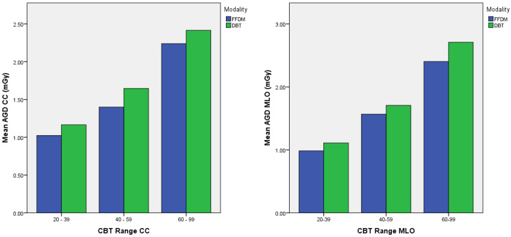
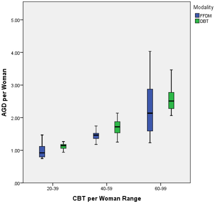
A set of national diagnostic reference levels (DRLs) was established in Malaysia for a range of breast thicknesses in 2013, but no updates for full-field digital mammography (FFDM) and digital breast tomosynthesis (DBT). Due to the increasing number of DBTs used and concern over radiation exposure, this study aimed to explore and establish local diagnostic reference levels for FFDM and DBT in Malaysia health facilities at different compressed breast thickness (CBT) ranges. The CBT, kilovoltage peak (kVp), Entrance surface dose (ESD), and average glandular dose (AGD) were retrospectively extracted from the mammography Digital Imaging and Communications in Medicine (DICOM) header. The 75th and 95th percentile values were obtained for the AGD distribution of each mammography projection for three sets of CBT range. The difference in AGD values between FFDM and DBT at three CBT ranges was determined. The DRLs for FFDM were 1.13 mGy, 1.52 mGy, and 2.87 mGy, while DBT were 1.18 mGy, 1.88 mGy, and 2.78 mGy at CBT ranges of 20−39 mm, 40−59 mm, and 60−99 mm, respectively. The AGD of DBT was significantly higher than FFDM for both mammographic views (p < 0.005). All three CBT groups showed a significant difference in AGD values for FFDM and DBT (p < 0.005). The local DRLs from this study were lower than the national DRLs, with the AGD of FFDM significantly lower than DBT.

摘要

2013年,马来西亚针对一系列乳房厚度制定了一套国家诊断参考水平(DRLs),但针对全场数字化乳腺摄影(FFDM)和数字乳腺断层合成(DBT)未进行更新。由于DBT的使用数量不断增加以及对辐射暴露的担忧,本研究旨在探索并确定马来西亚医疗机构中不同压缩乳房厚度(CBT)范围内FFDM和DBT的本地诊断参考水平。从乳腺摄影数字成像和通信医学(DICOM)头文件中回顾性提取CBT、峰值千伏(kVp)、体表入射剂量(ESD)和平均腺体剂量(AGD)。针对三组CBT范围,获取每个乳腺摄影投影AGD分布的第75百分位数和第95百分位数。确定了三个CBT范围内FFDM和DBT之间AGD值的差异。在CBT范围为20 - 39毫米、40 - 59毫米和60 - 99毫米时,FFDM的DRLs分别为1.13毫戈瑞、1.52毫戈瑞和2.87毫戈瑞,而DBT的DRLs分别为1.18毫戈瑞、1.88毫戈瑞和2.78毫戈瑞。对于两种乳腺摄影视图,DBT的AGD均显著高于FFDM(p < 0.005)。所有三个CBT组在FFDM和DBT的AGD值上均存在显著差异(p < 0.005)。本研究中的本地DRLs低于国家DRLs,FFDM的AGD显著低于DBT。

https://cdn.ncbi.nlm.nih.gov/pmc/blobs/e7f6/9601326/35c62dc32b4b/healthcare-10-01917-g002.jpg
https://cdn.ncbi.nlm.nih.gov/pmc/blobs/e7f6/9601326/3fe3acf190d5/healthcare-10-01917-g001.jpg
https://cdn.ncbi.nlm.nih.gov/pmc/blobs/e7f6/9601326/35c62dc32b4b/healthcare-10-01917-g002.jpg
https://cdn.ncbi.nlm.nih.gov/pmc/blobs/e7f6/9601326/3fe3acf190d5/healthcare-10-01917-g001.jpg
https://cdn.ncbi.nlm.nih.gov/pmc/blobs/e7f6/9601326/35c62dc32b4b/healthcare-10-01917-g002.jpg

相似文献

1
Local Diagnostic Reference Levels for Full-Field Digital Mammography and Digital Breast Tomosynthesis in a Tertiary Hospital in Malaysia.马来西亚一家三级医院全视野数字乳腺摄影和数字乳腺断层合成的局部诊断参考水平
Healthcare (Basel). 2022 Sep 30;10(10):1917. doi: 10.3390/healthcare10101917.
2
Comparison of radiation doses between diagnostic full-field digital mammography (FFDM) and digital breast tomosynthesis (DBT): a clinical study.诊断全数字化乳腺摄影(FFDM)与数字乳腺断层合成(DBT)的辐射剂量比较:一项临床研究。
J Med Radiat Sci. 2020 Sep;67(3):185-192. doi: 10.1002/jmrs.405. Epub 2020 Jun 3.
3
Local diagnostic reference levels (LDRLs) for full-field digital mammography (FFDM) and digital breast tomosynthesis (DBT) procedures in Morocco.摩洛哥全数字化乳腺摄影(FFDM)和数字乳腺断层合成(DBT)的局部诊断参考水平(LDRL)。
J Med Imaging Radiat Sci. 2022 Jun;53(2):242-247. doi: 10.1016/j.jmir.2022.03.008. Epub 2022 Mar 29.
4
Radiation exposure of digital breast tomosynthesis using an antiscatter grid compared with full-field digital mammography.数字乳腺断层合成摄影与全数字化乳腺摄影的散射辐射比较。
Invest Radiol. 2015 Oct;50(10):679-85. doi: 10.1097/RLI.0000000000000168.
5
Assessment of local diagnostic reference levels (LDRLS) for full-field digital mammography (FFDM) and digital breast tomosynthesis (DBT) mammography in Tamil Nadu, India.印度泰米尔纳德邦全数字化乳腺摄影术(FFDM)和数字乳腺断层合成术(DBT)乳腺摄影的局部诊断参考水平(LDRLS)评估。
Radiat Prot Dosimetry. 2024 Nov 18;200(19):1926-1932. doi: 10.1093/rpd/ncae199.
6
The effect of different exposure parameters on radiation dose in digital mammography and digital breast tomosynthesis: A phantom study.不同曝光参数对数字乳腺摄影和数字乳腺断层合成辐射剂量的影响:一项体模研究。
Radiography (Lond). 2020 Aug;26(3):e129-e133. doi: 10.1016/j.radi.2019.12.004. Epub 2019 Dec 20.
7
Breast Radiation Dose With CESM Compared With 2D FFDM and 3D Tomosynthesis Mammography.对比二维全视野数字化乳腺摄影(2D FFDM)和三维乳腺断层合成摄影,乳腺增强合成钼靶(CESM)的乳腺辐射剂量
AJR Am J Roentgenol. 2017 Feb;208(2):362-372. doi: 10.2214/AJR.16.16743.
8
Mammography Diagnostic Reference Levels (DRLs) in Ghana.加纳的乳房 X 光摄影诊断参考水平(DRLs)。
Radiography (Lond). 2021 May;27(2):611-616. doi: 10.1016/j.radi.2020.11.022. Epub 2020 Dec 18.
9
Evaluation of average glandular dose (AGD) in screening and diagnostic digital mammography and digital breast tomosynthesis (DBT) towards establishing a reference dose range band (DRB): a developing country experience.评估在筛查和诊断数字乳腺摄影和数字乳腺断层合成(DBT)中的平均腺体剂量(AGD),以建立参考剂量范围带(DRB):发展中国家的经验。
J Radiol Prot. 2023 Jul 26;43(3). doi: 10.1088/1361-6498/ace833.
10
Breast Radiation Exposure of 3D Digital Breast Tomosynthesis Compared to Full-Field Digital Mammography in a Clinical Follow-Up Setting.在临床随访环境中,与全视野数字化乳腺摄影相比,3D数字乳腺断层合成的乳腺辐射暴露情况
Diagnostics (Basel). 2022 Feb 10;12(2):456. doi: 10.3390/diagnostics12020456.

引用本文的文献

1
Local Diagnostic Reference Levels in Digital Breast Tomosynthesis.数字乳腺断层合成中的局部诊断参考水平
J Med Radiat Sci. 2025 Sep;72(3):341-349. doi: 10.1002/jmrs.892. Epub 2025 Jun 25.
2
Establishing Diagnostic Reference Levels in Digital Mammography from Eight Mammography Units Using over 30,000 Images.利用超过30000张图像,从八个乳腺钼靶摄影设备中建立数字乳腺钼靶摄影的诊断参考水平。
Diagnostics (Basel). 2025 Mar 10;15(6):682. doi: 10.3390/diagnostics15060682.
3
Assessing the Organ Dose in Diagnostic Imaging with Digital Tomosynthesis System Using TLD100H Dosimeters.

本文引用的文献

1
Comparison of Mean Glandular Dose between Full-Field Digital Mammography and Digital Breast Tomosynthesis.全视野数字乳腺摄影与数字乳腺断层合成平均腺体剂量的比较。
Healthcare (Basel). 2021 Dec 19;9(12):1758. doi: 10.3390/healthcare9121758.
2
Mammography Diagnostic Reference Levels (DRLs) in Ghana.加纳的乳房 X 光摄影诊断参考水平(DRLs)。
Radiography (Lond). 2021 May;27(2):611-616. doi: 10.1016/j.radi.2020.11.022. Epub 2020 Dec 18.
3
Comparison of radiation doses between diagnostic full-field digital mammography (FFDM) and digital breast tomosynthesis (DBT): a clinical study.
使用TLD100H剂量计评估数字断层合成系统在诊断成像中的器官剂量。
Tomography. 2025 Mar 11;11(3):32. doi: 10.3390/tomography11030032.
4
Lifetime Attributable Risk in Mammography Screenings in Dubai: The Influence of Breast Thickness and Age on Radiation Exposure.迪拜乳腺钼靶筛查中的终生归因风险:乳房厚度和年龄对辐射暴露的影响。
Diagnostics (Basel). 2025 Jan 2;15(1):83. doi: 10.3390/diagnostics15010083.
5
National diagnostic reference levels for digital diagnostic and screening mammography in Uganda.乌干达数字诊断和筛查乳房 X 光摄影的国家诊断参考水平。
PLoS One. 2024 Aug 29;19(8):e0294541. doi: 10.1371/journal.pone.0294541. eCollection 2024.
6
Screening Mammography Diagnostic Reference Level System According to Compressed Breast Thickness: Dubai Health.根据乳房压缩厚度的乳腺钼靶筛查诊断参考水平系统:迪拜健康局
J Imaging. 2024 Aug 5;10(8):188. doi: 10.3390/jimaging10080188.
7
Service Quality and Related Factors in Primary Health Care Services: A Cross-Sectional Study.基层医疗服务中的服务质量及相关因素:一项横断面研究
Healthcare (Basel). 2024 May 8;12(10):965. doi: 10.3390/healthcare12100965.
8
Demonstration of Japanese radiographic examination codes in establishing typical values for a wide variety of general radiography examinations.日本放射检查代码在为各种普通放射检查确定典型值方面的示范。
Sci Rep. 2024 Jan 26;14(1):2249. doi: 10.1038/s41598-024-52294-y.
9
Estimating Local Diagnostic Reference Levels for Mammography in Dubai.估算迪拜乳腺钼靶摄影的局部诊断参考水平。
Diagnostics (Basel). 2023 Dec 20;14(1):8. doi: 10.3390/diagnostics14010008.
诊断全数字化乳腺摄影(FFDM)与数字乳腺断层合成(DBT)的辐射剂量比较:一项临床研究。
J Med Radiat Sci. 2020 Sep;67(3):185-192. doi: 10.1002/jmrs.405. Epub 2020 Jun 3.
4
Can Digital Breast Tomosynthesis Replace Full-Field Digital Mammography? A Multireader, Multicase Study of Wide-Angle Tomosynthesis.数字乳腺断层合成能否取代全视野数字乳腺摄影?广角断层合成的多读者、多病例研究。
AJR Am J Roentgenol. 2019 Jun;212(6):1393-1399. doi: 10.2214/AJR.18.20294. Epub 2019 Apr 1.
5
INSTITUTIONAL BREAST DOSES IN DIGITAL MAMMOGRAPHY.数字化乳腺摄影中的机构乳腺剂量
Radiat Prot Dosimetry. 2019 Dec 23;185(2):239-251. doi: 10.1093/rpd/ncz005.
6
ICRP Publication 135: Diagnostic Reference Levels in Medical Imaging.国际放射防护委员会第135号出版物:医学成像中的诊断参考水平
Ann ICRP. 2017 Oct;46(1):1-144. doi: 10.1177/0146645317717209.
7
Radiation dose with digital breast tomosynthesis compared to digital mammography: per-view analysis.数字乳腺断层合成与数字乳腺钼靶摄影的放射剂量比较:单视图分析。
Eur Radiol. 2018 Feb;28(2):573-581. doi: 10.1007/s00330-017-5024-4. Epub 2017 Aug 17.
8
Mean glandular dose in digital mammography: a dose calculation method comparison.数字化乳腺摄影中的平均腺体剂量:剂量计算方法比较
J Med Imaging (Bellingham). 2017 Jan;4(1):013502. doi: 10.1117/1.JMI.4.1.013502. Epub 2017 Jan 24.
9
Diagnostic reference levels for digital mammography in New South Wales.新南威尔士州数字乳腺摄影的诊断参考水平
J Med Imaging Radiat Oncol. 2017 Feb;61(1):48-57. doi: 10.1111/1754-9485.12540. Epub 2016 Oct 6.
10
Digital breast tomosynthesis (DBT): a review of the evidence for use as a screening tool.数字乳腺断层合成(DBT):用作筛查工具的证据综述。
Clin Radiol. 2016 Feb;71(2):141-50. doi: 10.1016/j.crad.2015.11.008. Epub 2015 Dec 23.