Biomedical Image Analysis Group, Department of Computing, Imperial College London, London, UK.
Institute of Signal Processing and System Theory, University of Stuttgart, Stuttgart, Germany.
Sci Rep. 2022 Nov 4;12(1):18733. doi: 10.1038/s41598-022-23632-9.
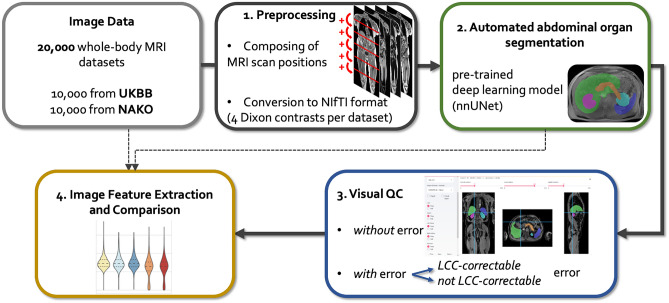
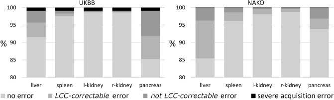
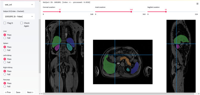
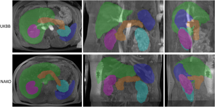
Large epidemiological studies such as the UK Biobank (UKBB) or German National Cohort (NAKO) provide unprecedented health-related data of the general population aiming to better understand determinants of health and disease. As part of these studies, Magnetic Resonance Imaging (MRI) is performed in a subset of participants allowing for phenotypical and functional characterization of different organ systems. Due to the large amount of imaging data, automated image analysis is required, which can be performed using deep learning methods, e. g. for automated organ segmentation. In this paper we describe a computational pipeline for automated segmentation of abdominal organs on MRI data from 20,000 participants of UKBB and NAKO and provide results of the quality control process. We found that approx. 90% of data sets showed no relevant segmentation errors while relevant errors occurred in a varying proportion of data sets depending on the organ of interest. Image-derived features based on automated organ segmentations showed relevant deviations of varying degree in the presence of segmentation errors. These results show that large-scale, deep learning-based abdominal organ segmentation on MRI data is feasible with overall high accuracy, but visual quality control remains an important step ensuring the validity of down-stream analyses in large epidemiological imaging studies.
大型流行病学研究,如英国生物银行(UKBB)或德国国家队列研究(NAKO),提供了前所未有的与健康相关的一般人群数据,旨在更好地了解健康和疾病的决定因素。作为这些研究的一部分,对一部分参与者进行磁共振成像(MRI)检查,以对不同器官系统进行表型和功能特征分析。由于成像数据量巨大,需要进行自动图像分析,这可以使用深度学习方法来完成,例如用于自动器官分割。本文描述了一个用于 UKBB 和 NAKO 中 20000 名参与者的 MRI 数据的自动腹部器官分割的计算流程,并提供了质量控制过程的结果。我们发现,大约 90%的数据集中没有出现相关的分割错误,而在不同的感兴趣器官中,数据集中会出现不同比例的相关错误。基于自动器官分割的图像衍生特征在存在分割错误的情况下表现出不同程度的显著偏差。这些结果表明,基于深度学习的大型 MRI 数据的腹部器官自动分割具有整体高精度,但视觉质量控制仍然是一个重要步骤,可确保在大型流行病学成像研究中下游分析的有效性。