Georgiev Stefan, Ruiss Manuel, Dana-Fisus Andreea, Leitgeb Rainer A, Findl Oliver
VIROS-Vienna Institute for Research in Ocular Surgery, A Karl Landsteiner Institute, Hanusch Hospital, Heinrich-Collin-Strasse 30, 1140, Vienna, Austria.
Center for Medical Physics and Biomedical Engineering, Vienna, Austria.
Eye Vis (Lond). 2023 Aug 1;10(1):30. doi: 10.1186/s40662-023-00348-z.
To comprehensively evaluate the agreement of component corneal aberrations from the newly updated wavefront analysis software of a swept-source optical coherence tomographer (SS-OCT) and a referential Placido-topography combined OCT device in elderly cataract patients.
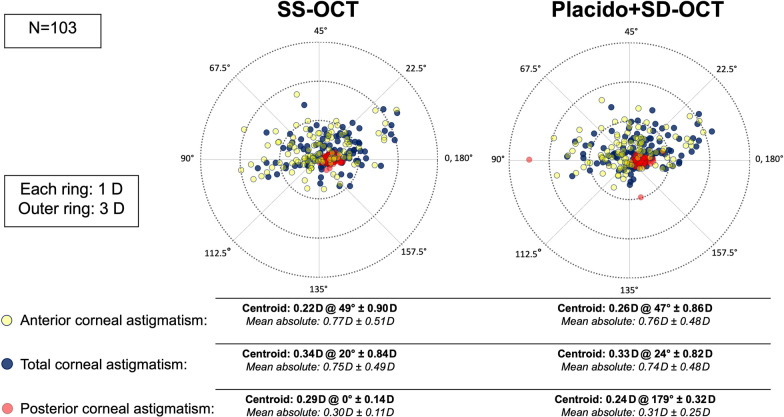
Retrospective study including 103 eyes from 103 elderly patients scheduled for cataract surgery that were measured on the same day with a SS-OCT (Heidelberg Engineering, Germany) device and a Placido-topography combined OCT device (CSO, Italy). Anterior, total, and posterior corneal wavefront aberrations were evaluated for their mean differences and limits of agreement (LoA) via Bland-Altman plots. Vector analysis was additionally employed to compare corneal astigmatism measurements in dioptric vector space.
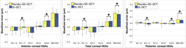
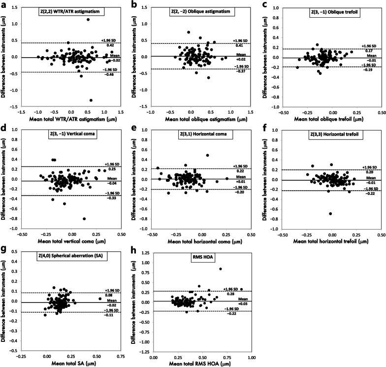
Mean differences of all corneal aberrometric parameters did not exceed 0.05 μm. Total corneal aberrations were not significantly different from 0 except for vertical coma (- 0.04 μm; P = 0.003), spherical aberration (- 0.01 μm, P < 0.001), and root mean square (RMS) higher-order aberration (HOA) (0.03 μm, P = 0.04). The 95% LoA for total corneal aberration parameters between both devices were - 0.46 to 0.42 μm for horizontal astigmatism, - 0.37 to 0.41 μm for oblique astigmatism, - 0.19 to 0.17 μm for oblique trefoil, - 0.33 to 0.25 μm for vertical coma, - 0.20 to 0.22 μm for horizontal coma, - 0.22 to 0.20 μm for horizontal trefoil, - 0.11 to 0.08 μm for spherical aberration, and - 0.22 to 0.28 μm for RMS HOA. Vector analysis revealed no statistically significant mean differences for anterior, total, and posterior corneal astigmatism in dioptric vector space.
In eyes undergoing cataract surgery with a regular elderly cornea, corneal wavefront analysis from the SS-OCT device showed functional equivalency to the reference device. Nevertheless, clinically relevant higher order aberration parameters should be interpreted with caution for surgical decision-making.
全面评估新型更新的扫频光学相干断层扫描仪(SS-OCT)波前分析软件与参考性角膜地形图联合OCT设备在老年白内障患者中角膜像差分量的一致性。
回顾性研究,纳入103例计划行白内障手术的老年患者的103只眼,于同一天使用德国海德堡工程公司的SS-OCT设备和意大利CSO公司的角膜地形图联合OCT设备进行测量。通过布兰德-奥特曼图评估前、全和后角膜波前像差的平均差异和一致性界限(LoA)。另外采用矢量分析在屈光度矢量空间比较角膜散光测量值。
所有角膜像差参数的平均差异均未超过0.05μm。除垂直慧差(-0.04μm;P = 0.003)、球差(-0.01μm,P < 0.001)和均方根(RMS)高阶像差(HOA)(0.03μm,P = 0.04)外,全角膜像差与0无显著差异。两种设备之间全角膜像差参数的95% LoA为:水平散光-0.46至0.42μm,斜向散光-0.37至0.41μm,斜向三叶像差-0.19至0.17μm,垂直慧差-0.33至0.25μm,水平慧差-0.20至0.22μm,水平三叶像差-0.22至0.20μm,球差-0.11至0.08μm,RMS HOA-0.22至0.28μm。矢量分析显示在屈光度矢量空间中前、全和后角膜散光无统计学显著平均差异。
在具有规则老年角膜的白内障手术眼中,SS-OCT设备的角膜波前分析显示与参考设备功能等效。然而,对于手术决策,临床相关的高阶像差参数应谨慎解读。