• 文献检索
  • 文档翻译
  • 深度研究
  • 学术资讯
  • Suppr Zotero 插件Zotero 插件
  • 邀请有礼
  • 套餐&价格
  • 历史记录
应用&插件
Suppr Zotero 插件Zotero 插件浏览器插件Mac 客户端Windows 客户端微信小程序
定价
高级版会员购买积分包购买API积分包
服务
文献检索文档翻译深度研究API 文档MCP 服务
关于我们
关于 Suppr公司介绍联系我们用户协议隐私条款
关注我们

Suppr 超能文献

核心技术专利:CN118964589B侵权必究
粤ICP备2023148730 号-1Suppr @ 2026

文献检索

告别复杂PubMed语法,用中文像聊天一样搜索,搜遍4000万医学文献。AI智能推荐,让科研检索更轻松。

立即免费搜索

文件翻译

保留排版,准确专业,支持PDF/Word/PPT等文件格式,支持 12+语言互译。

免费翻译文档

深度研究

AI帮你快速写综述,25分钟生成高质量综述,智能提取关键信息,辅助科研写作。

立即免费体验

基于细胞边界图像生成用于高分辨率空间转录组学的单细胞基因表达谱。

Generating single-cell gene expression profiles for high-resolution spatial transcriptomics based on cell boundary images.

作者信息

Zhang Bohan, Li Mei, Kang Qiang, Deng Zhonghan, Qin Hua, Su Kui, Feng Xiuwen, Chen Lichuan, Liu Huanlin, Fang Shuangsang, Zhang Yong, Li Yuxiang, Brix Susanne, Xu Xun

机构信息

BGI Research, Shenzhen, 518083, China.

BGI Research, Beijing, 102601, China.

出版信息

GigaByte. 2024 Feb 20;2024:gigabyte110. doi: 10.46471/gigabyte.110. eCollection 2024.

DOI:10.46471/gigabyte.110
PMID:38434932
原文链接:https://pmc.ncbi.nlm.nih.gov/articles/PMC10905256/
Abstract

UNLABELLED

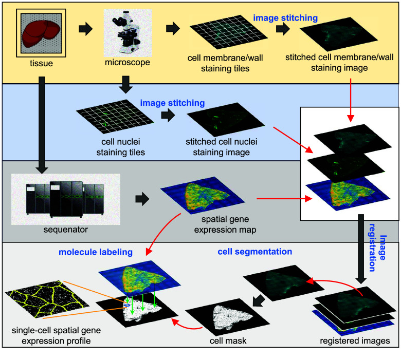
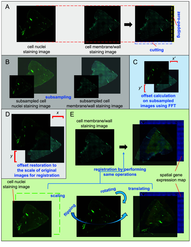
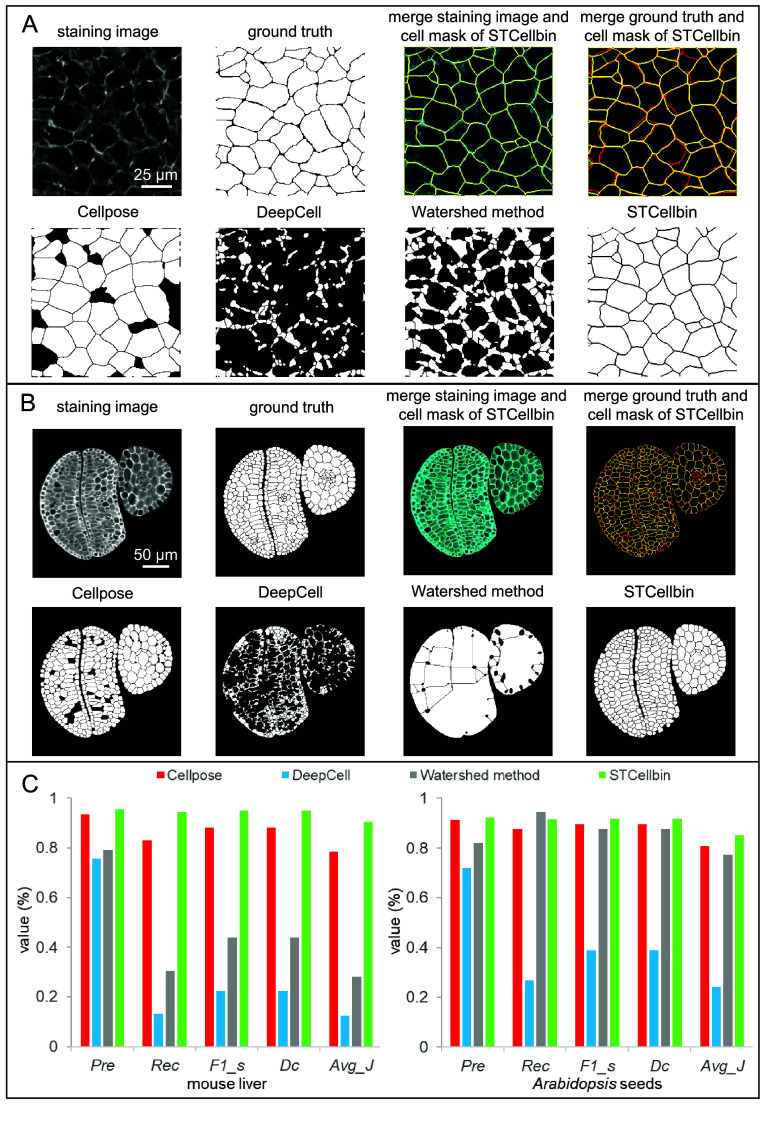
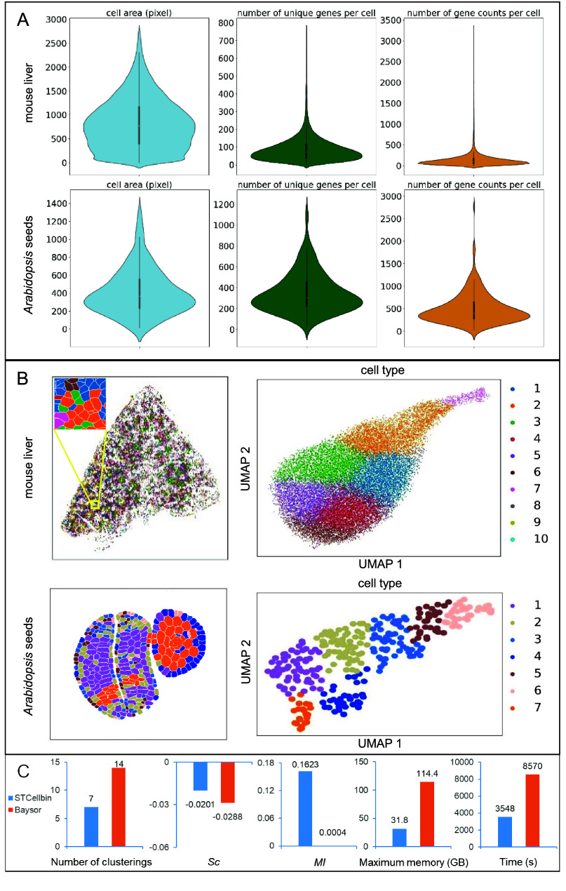
In spatially resolved transcriptomics, Stereo-seq facilitates the analysis of large tissues at the single-cell level, offering subcellular resolution and centimeter-level field-of-view. Our previous work on StereoCell introduced a one-stop software using cell nuclei staining images and statistical methods to generate high-confidence single-cell spatial gene expression profiles for Stereo-seq data. With advancements allowing the acquisition of cell boundary information, such as cell membrane/wall staining images, we updated our software to a new version, STCellbin. Using cell nuclei staining images, STCellbin aligns cell membrane/wall staining images with spatial gene expression maps. Advanced cell segmentation ensures the detection of accurate cell boundaries, leading to more reliable single-cell spatial gene expression profiles. We verified that STCellbin can be applied to mouse liver (cell membranes) and seed (cell walls) datasets, outperforming other methods. The improved capability of capturing single-cell gene expression profiles results in a deeper understanding of the contribution of single-cell phenotypes to tissue biology.

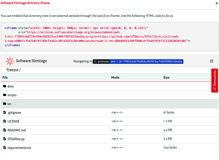
AVAILABILITY & IMPLEMENTATION: The source code of STCellbin is available at https://github.com/STOmics/STCellbin.

摘要

未标注

在空间分辨转录组学中,Stereo-seq有助于在单细胞水平上分析大型组织,提供亚细胞分辨率和厘米级视野。我们之前关于StereoCell的工作引入了一个一站式软件,该软件使用细胞核染色图像和统计方法为Stereo-seq数据生成高可信度的单细胞空间基因表达谱。随着能够获取细胞边界信息(如细胞膜/细胞壁染色图像)的技术进步,我们将软件更新到了新版本STCellbin。STCellbin利用细胞核染色图像将细胞膜/细胞壁染色图像与空间基因表达图谱对齐。先进的细胞分割确保了准确细胞边界的检测,从而产生更可靠的单细胞空间基因表达谱。我们验证了STCellbin可应用于小鼠肝脏(细胞膜)和种子(细胞壁)数据集,性能优于其他方法。捕获单细胞基因表达谱能力的提高有助于更深入地理解单细胞表型对组织生物学的贡献。

可用性与实现方式

STCellbin的源代码可在https://github.com/STOmics/STCellbin获取。

https://cdn.ncbi.nlm.nih.gov/pmc/blobs/3be0/10905256/dda3e5c1a5fc/gigabyte-2024-110-g005.jpg
https://cdn.ncbi.nlm.nih.gov/pmc/blobs/3be0/10905256/b2158727a5da/gigabyte-2024-110-g001.jpg
https://cdn.ncbi.nlm.nih.gov/pmc/blobs/3be0/10905256/bb422b60b21c/gigabyte-2024-110-g002.jpg
https://cdn.ncbi.nlm.nih.gov/pmc/blobs/3be0/10905256/b7e39461b811/gigabyte-2024-110-g003.jpg
https://cdn.ncbi.nlm.nih.gov/pmc/blobs/3be0/10905256/de0ee3f98cff/gigabyte-2024-110-g004.jpg
https://cdn.ncbi.nlm.nih.gov/pmc/blobs/3be0/10905256/dda3e5c1a5fc/gigabyte-2024-110-g005.jpg

相似文献

1
Generating single-cell gene expression profiles for high-resolution spatial transcriptomics based on cell boundary images.基于细胞边界图像生成用于高分辨率空间转录组学的单细胞基因表达谱。
GigaByte. 2024 Feb 20;2024:gigabyte110. doi: 10.46471/gigabyte.110. eCollection 2024.
2
Reference-free cell type deconvolution of multi-cellular pixel-resolution spatially resolved transcriptomics data.无参考细胞类型分解多细胞像素分辨率空间分辨转录组学数据。
Nat Commun. 2022 Apr 29;13(1):2339. doi: 10.1038/s41467-022-30033-z.
3
stPlus: a reference-based method for the accurate enhancement of spatial transcriptomics.stPlus:一种基于参考的方法,用于准确增强空间转录组学。
Bioinformatics. 2021 Jul 12;37(Suppl_1):i299-i307. doi: 10.1093/bioinformatics/btab298.
4
Integrating multi-modal information to detect spatial domains of spatial transcriptomics by graph attention network.通过图注意力网络整合多模态信息以检测空间转录组学的空间域。
J Genet Genomics. 2023 Sep;50(9):720-733. doi: 10.1016/j.jgg.2023.06.005. Epub 2023 Jun 23.
5
Computational solutions for spatial transcriptomics.空间转录组学的计算解决方案。
Comput Struct Biotechnol J. 2022 Sep 1;20:4870-4884. doi: 10.1016/j.csbj.2022.08.043. eCollection 2022.
6
SD2: spatially resolved transcriptomics deconvolution through integration of dropout and spatial information.SD2:通过整合缺失数据和空间信息进行空间分辨转录组学去卷积。
Bioinformatics. 2022 Oct 31;38(21):4878-4884. doi: 10.1093/bioinformatics/btac605.
7
EnDecon: cell type deconvolution of spatially resolved transcriptomics data via ensemble learning.EnDecon:通过集成学习对空间分辨转录组数据进行细胞类型去卷积。
Bioinformatics. 2023 Jan 1;39(1). doi: 10.1093/bioinformatics/btac825.
8
Single-cell level deconvolution, convolution, and clustering in spatial transcriptomics by aligning spot level transcriptome to nuclear morphology.通过将斑点水平转录组与核形态对齐,在空间转录组学中进行单细胞水平的反卷积、卷积和聚类。
bioRxiv. 2023 Dec 18:2023.12.17.572084. doi: 10.1101/2023.12.17.572084.
9
FISHFactor: a probabilistic factor model for spatial transcriptomics data with subcellular resolution.FISHFactor:具有亚细胞分辨率的空间转录组学数据的概率因子模型。
Bioinformatics. 2023 May 4;39(4). doi: 10.1093/bioinformatics/btad183.
10
iSC.MEB: an R package for multi-sample spatial clustering analysis of spatial transcriptomics data.iSC.MEB:一个用于空间转录组学数据多样本空间聚类分析的R包。
Bioinform Adv. 2023 Feb 17;3(1):vbad019. doi: 10.1093/bioadv/vbad019. eCollection 2023.

引用本文的文献

1
Single-Cell and Spatial Transcriptomic Profiling of Penile Squamous Cell Carcinoma Reveals Dynamics of Tumor Differentiation and Immune Microenvironment.阴茎鳞状细胞癌的单细胞和空间转录组分析揭示肿瘤分化和免疫微环境的动态变化
Adv Sci (Weinh). 2025 Sep;12(33):e00216. doi: 10.1002/advs.202500216. Epub 2025 Jun 5.
2
Integrative single-cell and spatial transcriptome analysis reveals heterogeneity of human liver progenitor cells.整合单细胞和空间转录组分析揭示了人类肝祖细胞的异质性。
Hepatol Commun. 2025 Feb 26;9(3). doi: 10.1097/HC9.0000000000000662. eCollection 2025 Mar 1.
3
Spatially Resolved Multiomics: Data Analysis from Monoomics to Multiomics.

本文引用的文献

1
High resolution mapping of the tumor microenvironment using integrated single-cell, spatial and in situ analysis.利用集成的单细胞、空间和原位分析技术对肿瘤微环境进行高分辨率图谱绘制。
Nat Commun. 2023 Dec 19;14(1):8353. doi: 10.1038/s41467-023-43458-x.
2
Review of research on the instance segmentation of cell images.细胞图像实例分割研究综述。
Comput Methods Programs Biomed. 2022 Dec;227:107211. doi: 10.1016/j.cmpb.2022.107211. Epub 2022 Oct 31.
3
Cellpose 2.0: how to train your own model.Cellpose 2.0:如何训练自己的模型。
空间分辨多组学:从单组学到多组学的数据分析
BME Front. 2024 Jan 13;6:0084. doi: 10.34133/bmef.0084. eCollection 2025.
Nat Methods. 2022 Dec;19(12):1634-1641. doi: 10.1038/s41592-022-01663-4. Epub 2022 Nov 7.
4
Spatially resolved epigenomic profiling of single cells in complex tissues.单细胞在复杂组织中的空间分辨表观基因组分析。
Cell. 2022 Nov 10;185(23):4448-4464.e17. doi: 10.1016/j.cell.2022.09.035. Epub 2022 Oct 21.
5
Computational Approaches and Challenges in Spatial Transcriptomics.计算方法与空间转录组学的挑战
Genomics Proteomics Bioinformatics. 2023 Feb;21(1):24-47. doi: 10.1016/j.gpb.2022.10.001. Epub 2022 Oct 14.
6
High-plex imaging of RNA and proteins at subcellular resolution in fixed tissue by spatial molecular imaging.通过空间分子成像在固定组织中以亚细胞分辨率对RNA和蛋白质进行高多重成像。
Nat Biotechnol. 2022 Dec;40(12):1794-1806. doi: 10.1038/s41587-022-01483-z. Epub 2022 Oct 6.
7
Spatially resolved clonal copy number alterations in benign and malignant tissue.良性和恶性组织中空间分辨的克隆拷贝数改变。
Nature. 2022 Aug;608(7922):360-367. doi: 10.1038/s41586-022-05023-2. Epub 2022 Aug 10.
8
Spatiotemporal transcriptomic atlas of mouse organogenesis using DNA nanoball-patterned arrays.使用DNA纳米球图案化阵列构建的小鼠器官发生时空转录组图谱。
Cell. 2022 May 12;185(10):1777-1792.e21. doi: 10.1016/j.cell.2022.04.003. Epub 2022 May 4.
9
Whole-cell segmentation of tissue images with human-level performance using large-scale data annotation and deep learning.使用大规模数据标注和深度学习实现具有人类水平性能的组织图像全细胞分割。
Nat Biotechnol. 2022 Apr;40(4):555-565. doi: 10.1038/s41587-021-01094-0. Epub 2021 Nov 18.
10
Cell segmentation in imaging-based spatial transcriptomics.基于成像的空间转录组学中的细胞分割
Nat Biotechnol. 2022 Mar;40(3):345-354. doi: 10.1038/s41587-021-01044-w. Epub 2021 Oct 14.