Department of Computer Science & Engineering, Indian Institute of Technology Patna, Patna, 801106, India.
Maharaja Surajmal Institute of Technology, Delhi, India.
Sci Rep. 2024 May 29;14(1):12380. doi: 10.1038/s41598-024-60861-6.
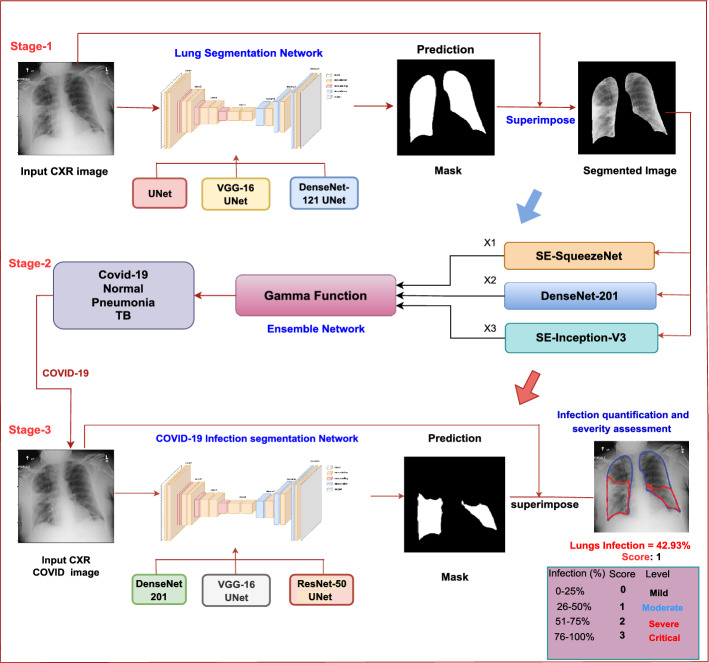
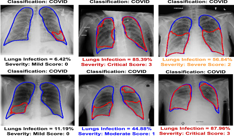
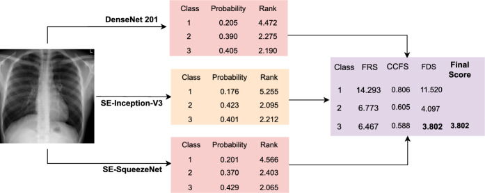
Chest Radiography is a non-invasive imaging modality for diagnosing and managing chronic lung disorders, encompassing conditions such as pneumonia, tuberculosis, and COVID-19. While it is crucial for disease localization and severity assessment, existing computer-aided diagnosis (CAD) systems primarily focus on classification tasks, often overlooking these aspects. Additionally, prevalent approaches rely on class activation or saliency maps, providing only a rough localization. This research endeavors to address these limitations by proposing a comprehensive multi-stage framework. Initially, the framework identifies relevant lung areas by filtering out extraneous regions. Subsequently, an advanced fuzzy-based ensemble approach is employed to categorize images into specific classes. In the final stage, the framework identifies infected areas and quantifies the extent of infection in COVID-19 cases, assigning severity scores ranging from 0 to 3 based on the infection's severity. Specifically, COVID-19 images are classified into distinct severity levels, such as mild, moderate, severe, and critical, determined by the modified RALE scoring system. The study utilizes publicly available datasets, surpassing previous state-of-the-art works. Incorporating lung segmentation into the proposed ensemble-based classification approach enhances the overall classification process. This solution can be a valuable alternative for clinicians and radiologists, serving as a secondary reader for chest X-rays, reducing reporting turnaround times, aiding clinical decision-making, and alleviating the workload on hospital staff.
胸部 X 光摄影是一种用于诊断和管理慢性肺部疾病的非侵入性成像方式,涵盖了肺炎、肺结核和 COVID-19 等疾病。虽然它对于疾病定位和严重程度评估至关重要,但现有的计算机辅助诊断 (CAD) 系统主要侧重于分类任务,往往忽略了这些方面。此外,流行的方法依赖于类激活或显着性映射,仅提供大致的定位。本研究通过提出一个全面的多阶段框架来解决这些限制。首先,该框架通过过滤掉无关区域来识别相关的肺部区域。然后,采用先进的基于模糊的集成方法将图像分类为特定的类别。在最后阶段,该框架识别感染区域,并量化 COVID-19 病例中的感染程度,根据感染的严重程度分配 0 到 3 的严重程度评分。具体来说,COVID-19 图像根据改良的 RALE 评分系统分为不同的严重程度级别,如轻度、中度、重度和危重度。该研究利用了公开可用的数据集,超过了以前的最先进的工作。将肺部分割纳入基于集成的分类方法中,增强了整体分类过程。该解决方案可以为临床医生和放射科医生提供有价值的替代方案,作为胸部 X 光的辅助读者,减少报告周转时间,辅助临床决策,并减轻医院工作人员的工作量。